2018 Winner’s Circle
This article is part of our FOCUS 2018 Annual Newsletter which will be published in full in March 2019.
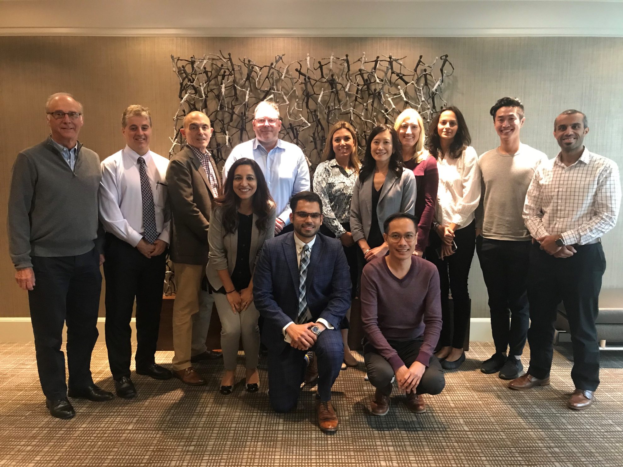
In each issue of the Foundation’s annual newsletter, titled FOCUS, the four highest-scoring resident and faculty researchers who submitted proposals in the prior year’s Spring and Fall cycles of the Competetive Research Grant Program are profiled. This gives our supporters a chance to learn more about the endodontists that received funding from the Foundation for Endodontics. The top-scoring resident and faculty researchers receive a $500 personal award in addition to having their projects selected for funding by the AAE’s Research and Scientific Affairs Committee, pictured below.
SPRING 2018 Faculty Winner
 Jennifer L. Gibbs, MAS, D.D.S., Ph.D.
Harvard School of Dental Medicine
Project: Identification of Clinical and Biological Risk Factors for Pain After Root End Resection Surgery
Endodontists are experts in pain management and Dr. Jennifer L. Gibbs is on a mission to keep it that way. Her project, titled, Identification of Clinical and Biological Risk Factors for Pain After Root End Resection Surgery, seeks to identify risk factors for pain in post-operative patient experiences. Her study, funded by the Foundation for Endodontics in the spring of 2018, is driven by Gibbs’ belief in the need for endodontists to have more specific information about what drives post-operative pain.
Pain management is essential to success in endodontics. Many patients’ first question before surgery is, “How much will this hurt?” If equipped with knowledge of specific risk factors that contribute to pain, clinicians might be better equipped to identify those patients who will need extra care and intervention in order to have a successful surgical recovery period.
Inspired to use surgery as a model to study risk factors for pain, Gibbs hopes to make sense of the incredible variability she has found in her data so far. When asked about the perceived average pain experience, she said, “It’s really striking how what we might call the ‘average’ pain experience after surgery is simply not the reality for many patients. I’m finding that patient factors seem to drive the post-op pain experience as much or even more than specific surgical factors”
Dr. Gibbs will soon be transitioning to a program director role at Harvard University’s Department of Endodontics. In addition to adding to the available information on risk factors for pain after surgery, Gibbs also believes there is still much to learn about dental pulp and how it relates to repair and regeneration in addition to general health and disease.
Dr. Gibbs has been enjoying her family’s transition to life in Boston after moving from New York City in August. She is excited about an upcoming ski trip in Vermont.
SPRING 2018 Resident Winner
 Fang-Chi Li, D.D.S., Ph.D.
University of Toronto
Project: Nanoparticle Guided Dentin Micro-Tissue Engineering: Characterizing Fluid Dynamics and Mechanical Characteristics
Dr. Fang-Chi Li has always had deep respect for her chosen field of endodontics. She says her interest in endodontics stems from her wish to utilize engineering principles to surmount practical frustrations and roadblocks faced by clinicians. “Endodontic treatment has long been the treatment of choice to maintain the long-term functional requirements of a natural tooth, but even when the highest standards of clinical procedures are followed, microbial biofilms still persist within the anatomic complexities and uninstrumented portions of the root canal system.” Dr. Li’s research project, titled Nanoparticle Guided Dentin Micro-Tissue Engineering: Characterizing Fluid Dynamics and Mechanical Characteristics seeks to make progress towards developing a minimal invasive treatment strategy with dual objectives of improving the antibacterial efficacy within root canal system and enhancing the mechanical integrity of remaining dentin against vertical root fracture. She hopes doing so will enhance predictability and improve root canal efficiency.
Dr. Li’s in vitro study will focus on developing a strategy to deliver the nanoparticles efficiently to root canal dentin using a novel activated-microbubble method. The findings will provide an insight of fluid dynamics associated with MB strategy and to determine a way to apply nanoparticles into root canal system practically.
Working to incorporate micro-tissue principles with nanotechnology with Dr. Anil Kishen in the labs at the University of Toronto has been exciting, she says. “Overall, this nanoparticle-based application has the potential to provide a minimal invasive way to treat the infected teeth and enhance the longevity of endodontically treated teeth and this may be the next revolution in our field.”
Dr. Li says that the more she studies and researches in endodontics, the more she appreciates our natural structures and systems and how they function concordantly. In her free time, Dr. Li enjoys practicing piano, exercising in the gym and participating in church activities. She also loves to bring a laptop to her favorite corner coffee shop on a snowy day.
FALL 2018 Faculty Winner
 Leticia Chaves de Souza, D.D.S., M.S., Ph.D.
University of Texas Health Science Center at Houston
Project: Evaluation of the Physical-Chemical and Biological Properties of a New Bioceramic Sealer
In the fall of 2018, the Foundation for Endodontics funded Dr. Leticia Chaves de Souza’s research project, titled, Evaluation of the Physical-Chemical and Biological Properties of a New Bioceramic Sealer. In her study, Dr. Chaves de Souza hopes to provide useful data for clinicians to employ in their daily practice when deciding which endodontic materials to use in the many varied cases they encounter.
The investigation of the properties of endodontic materials is quite important. Dr. Chaves de Souza believes that as endodontists, it is necessary to fully understand the physical-chemical and biological properties of endodontic sealers, so that the best suitable materials may be chosen for each case. After the most appropriate sealer has been chosen, Dr. Chaves de Souza emphasizes that the proper obturation technique be understood for each sealer.
When asked what motivated her desire to investigate endodontic materials’ properties, Dr. Chaves de Souza explained, “The properties of these materials directly influence their clinical behavior as well as their handling characteristics, both of which will ultimately guide the clinical decisions made by endodontists.”
Dr. Chaves de Souza enjoys working with dental students and residents. She is interested in physical-chemical and biological properties of endodontic materials, endodontic instruments and understanding the molecular mechanisms underlying apical periodontitis. In her free time, Dr. Chaves de Souza enjoys going to the movies and travelling with family and friends.
Fall 2018 Resident Winner
 Omid Seyed Dianat, D.D.S., M.S.
University of Maryland
Project: The Accuracy and Efficiency of a Dynamic 3D Navigation System for Negotiating Calcified Canals
Dr. Omid Seyed Dianat is a second-year endodontics resident at the University of Maryland. His study, funded by the Foundation in the fall of 2018, titled The Accuracy and Efficiency of a Dynamic 3D Navigation System for Negotiating Calcified Canals, examines the accuracy and precision of dynamic navigation in endodontics.
Root canal configuration and morphology creates many challenges in everyday endodontic practice. One such challenge is calcification following restorative procedures and traumatic injuries. Despite breakthroughs in technology, canal localization in calcified canals is still considered to be a time consuming and stress-inducing procedure for clinicians.
In recent years, static guides and templates have been used to address and manage this issue with some degree of success and many limitations. There is a new computer-based technology system called X-Guide, which is currently being used for guided implant placement. X-Guide is a 3D navigation technique developed by X-Nav technologies that allows clinicians to use cone beam computed tomography (CBCT) to plan and place implants with great precision. Dr. Dianat believes the system could be adapted for endodontic practice and aid endodontists in locating calcified canals. In his study, Dr. Dianat hypothesizes that this method using X-Guide is accurate, saves time, results in more successful attempts to located canals and will overcome the limitations of static guides.
The findings of Dr. Dianat’s study will provide an understanding of the limitations of the X-Guide technology while gathering information for its clinical applications and future investigations.
Dr. Dianat is grateful for the family and friends who have supported him through his studies. He feels especially thankful for all the academic mentors he’s had while pursuing a career in endodontics, both at the University of Maryland and previously at Shahid Beheshti and Isfahan Universities of Medical Sciences in Iran. In his free time, Dr. Dianat enjoys spending time with family, swimming, biking, traveling and exploring other parts of the world.