
Enhancing Dental Education: Enabling Student Self-Evaluation in Endodontic Access Cavity Preparation Using 3D Software
By Ane Poly, DDS, MSc, PhD
As we navigate the rapidly evolving landscape of dental education, exploring how digital technologies can enhance current teaching methods and improve student learning is paramount. Endodontic access preparation (EAP) represents the critical first phase in non-surgical root canal treatment. Its primary objectives, such as conserving sound tooth structure, unroofing the pulp chamber, and establishing direct-line access to canal orifices, lay the foundation for clinical success.
Traditionally, preclinical courses rely on visual inspection by calibrated faculty members to grade student EAP. However, extensive literature shows that visual inspection is inherently subjective, leading to a lack of interrater reliability among faculty in the evaluation process.
Standard visual inspection also leaves students with limited, qualitative feedback. While an instructor can observe that an access cavity is overextended or off-center, students often struggle to visualize the exact three-dimensional volumetric defect or millimeter deviations from ideal dimensions. Because self-assessment is a crucial lifelong skill predoctoral students must develop to refine their hand-skills and work independently, establishing objective standards for error detection is essential.
Adapting 3D Comparison Software for Endodontics
To bridge this gap, CAD/CAM technology widely used in fixed prosthodontics to evaluate crown preparations (Planmeca Romexis Compare, formerly E4D Compare) was adapted for endodontic education to evaluate student EAPs three-dimensionally against standardized ideal master models.1
Students start by using intraoral scanners to scan their EAPs and upload the 3D file into the software. Then, they trace the access perimeter as an “Axial Base” and follow the board licensure landmarks (ADEX criteria2) to trace the “Margin”. High-resolution surface scanning superimposes the student model onto a faculty-determined ideal model, and the existing software tools are used to measure internal wall dimensions and outline extents:
- Compute Difference: Quantifies volumetric alignment via 3D heat maps, highlighting overextended areas in red, underextended areas in blue, and acceptable areas in yellow. (Figure 1)
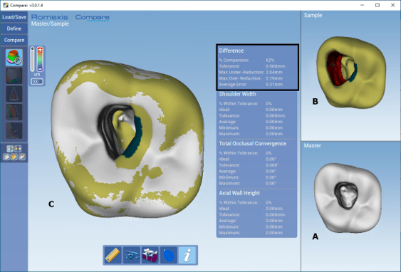
Figure 1: Compute difference tool showing (A) the ideal model, (B) the S2 model, and (C) the two models overlapped. Red areas indicate overextension; blue areas indicate underextension; yellow areas fall within the tolerance range. The black rectangle highlights the quantitative data.
- Slice Plane & Distance Tools: Provide cross-sectional views at various depth levels, enabling specific millimeter measurements between internal preparation walls. (Figure 2)
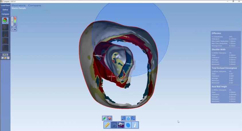
Figure 2: Ideal and student models overlapped with the slice plane tool showing a qualitative comparison of the internal form. Three measurements are shown in specific areas, illustrating the distance tool being used to measure the distance between models.
- Shoulder Width: Measures circumferential boundary distances to verify adherence to licensure safety margins.2 (Figure 3)
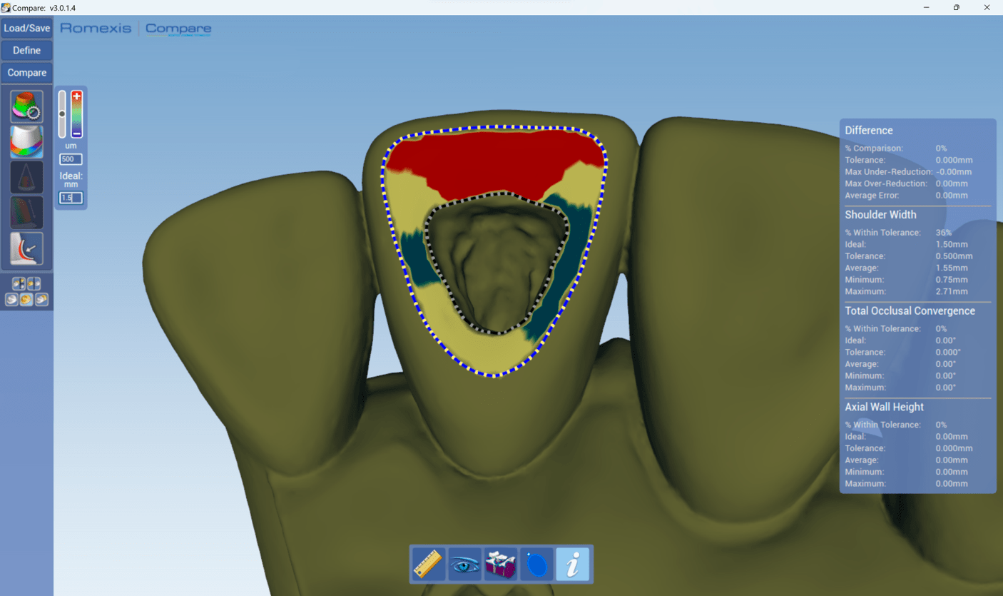
Figure 3: Shoulder width tool shows the student model where the red areas indicate the distance is > 2.0 mm, blue areas < 1.0 mm, and yellow areas between 1 and 2 mm.
Practical Insights from Preclinical Implementation
The viability and impact of this digital methodology were evaluated with a randomized controlled trial.3 Sixty second-year dental students performed EAPs on #14 acrylic teeth (RTE #14 With Insert; Acadental, Lenexa, KS) following the ADEX criteria2. Students were assigned either to a control group using traditional visual assessment or an experimental group combining traditional self-assessment with 3D software evaluation.
The trial revealed significant findings regarding student self-perception and critical evaluation. While both groups experienced an increase in clinical confidence, students relying solely on traditional visual assessment significantly overestimated their performance improvements. Conversely, students equipped with 3D digital feedback examined their work far more critically and accurately.
This outcome highlights the Dunning-Kruger effect, a cognitive bias where individuals with lower competence overestimate their abilities due to a lack of detailed feedback. The 3D comparison software serves as a “digital magnifying glass,” exposing minor over-extensions, wall gouging, and lack of straight-line access. By removing visual ambiguity, the software tempers unearned overconfidence and fosters genuine self-awareness.
Looking Ahead: Impact on Dental Curriculum and Faculty Workflow
Student acceptance has been positive. In our randomized trial, 96.7% of students who used the 3D method agreed that 3D evaluation should be permanently incorporated into preclinical endodontic training and licensure exam preparation.
Beyond student self-evaluation, integrating 3D evaluation software into preclinical simulation laboratory training helps address a broader institutional challenge: faculty shortages and calibration. With vacant budgeted faculty positions in U.S. dental schools reaching record highs, digital evaluation provides an objective, standardized baseline for instruction.
Ultimately, this precise and immediate feedback empowers students to critically analyze their work and build the lifelong self-assessment capabilities necessary for modern endodontic practice. Furthermore, by providing round-the-clock feedback, the software mitigates faculty workload, allowing instructors to redirect their time toward targeted, high-level clinical coaching.
Note: Generative AI was used as an editorial aid to assist with grammar, clarity, phrasing, and organization. The ideas, opinions, and final wording of the content are solely those of the author.
Ane Poly, DDS, MSc, PhD, is a Clinical Assistant Professor in the Department of Endodontics at the University of Florida College of Dentistry.
References
- Poly A, Burnett JE, Buie CA, Schweitzer JL. Three-dimensional software adapted to evaluate endodontic access cavity preparation. J Dent Educ. 2023;87(suppl 3):1848-1851.
- CDCA-WREB-CITA. ADEX Dental Exam Series: Endodontic Criteria. 2026 Candidate Manual. Anterior Endo Procedure: https://adextesting.org/wp-content/uploads/2026/05/2026.8-DEN-ENDO-Anterior_20260521.pdf Posterior Endo Procedure: https://adextesting.org/wp-content/uploads/2026/05/2026.8-DEN-ENDO-Posterior_20260521.pdf
- Poly A, Harness C, Vu E, Biradar A, Buie CA, Burnett JE, Schweitzer JL. Integrating digital technology in endodontic education: A randomized controlled trial evaluating student self-assessment and perspectives. J Dent Educ. 2025 Aug;89(8):1294-1302.
Disclaimer
The views and opinions expressed by authors are solely those of the authors and do not necessarily reflect the official policy or position of the American Association of Endodontists (AAE). Publication of these views does not imply endorsement by the AAE.