The Way We Were
 By Robert S. Roda, D.D.S., M.S.
“Progress is impossible without change, and those who cannot change their minds cannot change anything.” -George Bernard Shaw
I recall the first time I was terrified to do endodontic therapy.
I had worked as a general dentist for 10 years prior to entering my endodontic program and up until then, I had no fear of endodontics at all. That’s because of two things: first, when I was a general dentist, I had no idea what was really going on in endodontics, and second, after botching my very first two molar endo’s when first getting out of school, I referred all of my endodontic cases to the endodontist! No fear, no problem! But this was different. I was a resident. There was this faculty member in my grad program. He was tough, demanding, brilliant, and one of the best critical thinkers I have ever met. He was, at once, my father, and my superior officer and I had ultimate respect and ultimate love for that man, but sometimes he scared the heck out of me! He walked into my operatory one day, when I had no patient, and said “Rob, I find that I am in need of root canal therapy, and you are gonna do it for me.”
My blood ran cold. Not only was I to do it on a faculty member (this faculty member), but it was a heavily restored second molar and was very calcified to boot! Three angled radiographs (film that I hand dipped in a tank in my operatory to develop the image rapidly) did not show any evidence of canals, but the apical radiolucency was there. I had no confidence that I would be able to find the canals with my 1.5x power loupes (I remember wishing that someone would invent a light to attach to them). I had some Mueller burs, but they were pretty long and hard to use in molars, and the head of the contra-angle always blocked my vision. I assumed the tooth had three canals, since the literature said even first molars only had 4 about 25% of the time and I was concerned that my very stiff stainless steel hand files would be difficult to get around the significant curvatures in the roots.
Somehow, I managed to complete the procedure without event, but I found the canals using only feel and dumb luck by drilling holes in the chamber floor where I hoped the canals were. The working length radiographs showed me that my instruments were in the canal, and not the ligament (Whew!). After opening the canals with small K-files to #25 (the most likely hand instrument to separate, according to the current studies and also, about the largest instrument I could get around those curvatures), I flared them with pre-curved Hedstrom files using hand step-back filing. Once I had that done, I could relax. I just placed a cotton pellet with CMCP fumes in there and closed it with temporary restorative for the obligatory second visit a week later to obturate and see how badly I had transported the canals. The final case was actually great, but very stressful to do.
The only other time I was that stressed was a few months later when, as a newly minted second year resident, I was allowed to do my first apical surgery. In my program, first year residents had to be the primary chairside assistant for the surgeries done by the second years. I had assisted on 20-30 surgeries that year, I knew the literature cold, and I was ready for my first one. As it turned out, we got a new surgical device that year also. It was a way of doing apical preps without a slow speed microhandpiece and hand chisels. It was some type of device called an ultrasonic. It cut preps really well on extracted teeth, but now I was going to get to try it on a real case. The tooth was #30, and I was happy it was on my side, unlike #19 so I could see better. I was waiting for the local anesthesia when my Department Chair walked in with another man who was introduced to me as the President of a European country’s Endodontic Society. Apparently, not only was I about to do my first apical surgery with a brand-new apical preparation device (and it was on a molar), but I also had to narrate and describe the rationale for all of it to a visiting dignitary! I cut a full thickness flap using a 15C scalpel blade, reflected it using a periosteal elevator with undermining elevation, and then found the mental foramen and protected it throughout. I searched for the most likely spot to do my osteotomy by imagining where the roots went under the bone. I found the roots, resected them (with a bevel), and started my root end preparations. My loupes were so helpful in seeing what I was doing (I remember wishing that someone would invent a light to attach to them), and my junior resident (himself with many years of general practice behind him) did an amazing job of keeping everything dry and visible. I filled the root ends with Super-EBA (zinc-oxide mixed with ethoxybenzoic acid), the latest and greatest root-end filling material, and closed the flap with compression, chromic gut (no bacteria-sucking braided silk for me), and compression. Every step of the way I was narrating to the visiting dignitary, and that was the stressful part. For him, he loved it. He was so impressed with the almost magical nature of what we were doing here in America, and the fact that we would remove the sutures in 48 hours blew his mind. (It would be several years before the Internet would exist and really level the clinical playing field around the world.)
This is how clinical endodontics was done when I was a resident. It does not seem so long ago to me, and I don’t feel that old, but I know it must seem strange and primitive to you, the reader, 30 years later. My endodontic career has encompassed more rapid advances in my field than I could ever have imagined when I was a resident. It’s been like living in a science fiction novel. I recall the first time I looked at the histology of periradicular and pulpal healing after an early article about the first endodontic bioceramic, mineral trioxide aggregate (MTA) was published. Compared to everything that had existed before, it was like magic.
Electronic apex locators had been around in one form of another since 1961, but no matter how many I bought (and I bought a few) and how hard I tried to get them to work, they were just never as accurate as radiographic working length measurements. Then, along came the third-generation machines in the early 1990’s and all of a sudden, they were incredibly reliable. I am still using the units that I bought in 1994 in my clinic today!
Using nickel-titanium (NiTi) to make hand files had existed before I got into my residency, but they were, at that point, just another incremental modification like rhomboid file cross-section or 0.29 percent tip diameter increases between files that seemed to help some people, but never really made a big difference to me. Engine-driven instrumentation had its start in the 1970’s, but that had never become popular due to many technical limitations. It was the marriage of engine driven and NiTi instruments that suddenly changed everything. There were some early stumbles along the way, but the progression of the instrument design and improved metallurgy over the last three decades has not only revolutionized how we prepare canals, but for me personally, it extended my career by decades because it stopped the progression of carpal tunnel syndrome that I was developing due to all of that hand filing!
Microscopes literally opened my eyes and allowed me to see things that were absolutely invisible before. When I decided to leave general practice and go into endo (shortly after the launching of the Hubble Space Telescope, and as the Soviet Union crumbled), a periodontist friend of mine laughed when I told him I wanted to become an endodontist. “It’s the only specialty you can do when you are blindfolded.” he quipped! Well, after introduction of the dental operating microscope, that’s just not true! It has literally changed how endodontics is delivered. Now, first molars have 4th canals up to 93% of the time, and it’s not that they were not there before. The enhanced illumination and magnification have allowed creative members of the profession to develop new ways to handle all sorts of previously untreatable clinical situations. The other thing that the microscope did for me was to extend my career by decades since it put me in a much better ergonomic position and arrested the incipient back disorder I was developing!
And speaking of opening our eyes, let us not forget that use of small field of view CBCT is not that old either. It has transformed our ability to see the clinical situation better than ever before and has prevented untold numbers of needless exploratory procedures while allowing us to more predictably treat situations that were impossible before.
The pace of change will only become more rapid, I suspect and new concepts and technologies will continue to arrive in clinical practice that either allow for the profession to offer new capabilities to deal with still difficult clinical problems, or to improve the efficiency, safety, and outcomes of current therapy. The process of change does not always go smoothly, however. Some things appear to be very promising and well thought out, and yet they end up not fulfilling their promise. This is where science guides us, and while we are all excited about new developments, always test them with clinical trials to see if they actually work as envisioned, or whether the body of knowledge shows that they will not actually be useful. Or, you can do what I did and succumb to the marketing hype. I bought all the new stuff only to find that, with initial use, much of it was useless. You too can get a closet full of very expensive toys, that you accumulate over the years, that ends up being worthless junk. I wouldn’t recommend that approach, but if you like collecting dreck, then go for it!
As I approach the end of my career, I think about the challenges and wonders that lie ahead for the younger members of my profession. The challenges are obvious: burdensome student debt which limits choices of practice opportunities, finding stability in practice environments, and managing work/life balance, just to name a few. You can help to mitigate these and other challenges by stepping up and taking an active role in dental organizations like the AAE, but also take some joy in the wonders that will surely arrive to move the profession forward into the future. Guided endodontic surgery and canal finding are moving along well, as are methods that are developing to enhance the cleaning and disinfection of canals. Will robotic endodontic surgery ever happen? Will we ever find a way to prevent or predictably manage fractured teeth? To quote Rogers and Hammerstein, “What will the future bring? I wonder.”
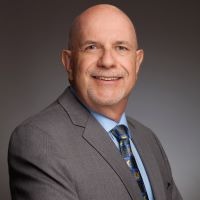
Dr. Robert S. Roda is a Past President of the AAE and a former Associate Editor for the Journal of Endodontics.


