
The AI Detective: Safeguarding Academic Integrity in Dental Education
 By Dr. Mohamed Ibrahim
Academic integrity is a cornerstone of dental education. In preclinical and clinical training, students are not only learning technical procedures but also developing the ethical habits that will guide their future patient care. As assessment methods continue to evolve, dental schools need reliable ways to ensure fairness, consistency, and trust in competency-based education.
In preclinical endodontic education, students are often assessed through nonsurgical root canal treatment procedures on typodont or 3D-printed teeth. Radiographs are taken at different stages of treatment, including the preoperative image, working length determination, master apical file, master apical cone, and final obturation. These images document treatment progression and help verify that the procedure was completed on the same tooth.
However, traditional oversight may not always detect subtle forms of academic dishonesty. For example, a student could potentially substitute a radiograph from another tooth or another procedure, making inconsistencies difficult to identify through manual review alone. While most students act with honesty and professionalism, even rare incidents can affect fairness and trust in the assessment process.
To address this challenge, our team developed an artificial intelligence model to detect inconsistencies among radiographs taken during preclinical endodontic procedures. The goal was not to replace faculty judgment, but to provide an additional objective screening tool to support educators in identifying cases that require closer review.
The model was based on a Siamese neural network, an AI architecture designed to compare two images and determine how similar they are. In this project, the model compared pairs of radiographs taken during different stages of root canal treatment. Radiographs from the same student’s procedure should demonstrate consistent tooth morphology and treatment progression, while images from different teeth or cases may show discrepancies.
The dataset included 3,390 radiographs from six previous preclinical nonsurgical root canal treatment competency exams involving 678 students. These radiographs allowed the model to evaluate continuity across treatment stages and determine whether submitted images appeared to belong to the same tooth.
The AI model showed strong performance. It achieved an overall accuracy of 89.31%, with precision of 76.82%, sensitivity of 84.82%, and an F1-score of 80.50%. The optimal similarity threshold was identified at 0.48. Cases close to this threshold were classified as “inconclusive,” meaning they required faculty review rather than an automatic decision. Figures 1 &2 show example of inconsistent pairs with low similarity scores.
This human oversight is essential. In academic integrity matters, fairness requires careful interpretation, context, and professional judgment. Therefore, any case flagged as inconsistent or inconclusive should be reviewed by faculty before a final determination is made. The AI system serves as a first layer of screening, helping educators focus attention on cases that may need further evaluation.
The study also examined how AI support influenced educator decision-making. Fifteen dental educators reviewed radiograph pairs during mock exam conditions. Without AI assistance, educators correctly identified 12.82% of manipulated radiographs. With AI guidance, detection accuracy increased to 63.89%. These findings suggest that AI can help educators identify subtle inconsistencies that may otherwise be missed.
At the same time, AI assistance increased review time. The average decision time increased from 143.15 seconds without AI to 263.92 seconds with AI. This likely reflects the additional effort required to interpret AI-generated labels and review flagged cases carefully. Although this adds cognitive load, it may be a reasonable trade-off when the goal is to improve accuracy, fairness, and confidence in high-stakes assessments.
The project also highlighted important lessons for responsible AI implementation. First, faculty training is critical. Educators need clear guidelines on how to interpret AI outputs, especially inconclusive results. Second, the use of AI should remain transparent and ethically grounded. Students should understand that the purpose of the system is to promote fairness and consistency, not to create a punitive environment. Third, AI outputs should be documented and reviewed through an established academic integrity process.
The model has limitations. Its performance depends on standardized radiographic imaging. Variations in exposure, angulation, or image quality may affect classification and lead to false-positive or inconclusive results. In addition, the current model was developed using data from a single institution. Future studies should validate the approach across multiple dental schools to determine whether the model performs consistently in different educational settings.
Despite these limitations, this work demonstrates the potential of artificial intelligence to strengthen assessment integrity in dental education. By identifying radiographic inconsistencies more efficiently and objectively, AI can support faculty while preserving fairness for students. More broadly, this approach shows how AI can be used responsibly in education: not as a replacement for educators, but as a tool that supports better decision-making.
As dental education continues to incorporate new technologies, the focus should remain on trust, transparency, and ethical implementation. Responsible AI integration can help promote academic integrity, support faculty, and prepare students for a profession where honesty, accountability, and technical excellence are inseparable.
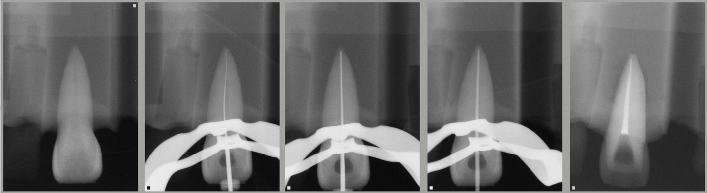
Figure (1) Preoperative and obturation inconsistent pairs with low similarity scores 0.003, representing radiographs from different cases.
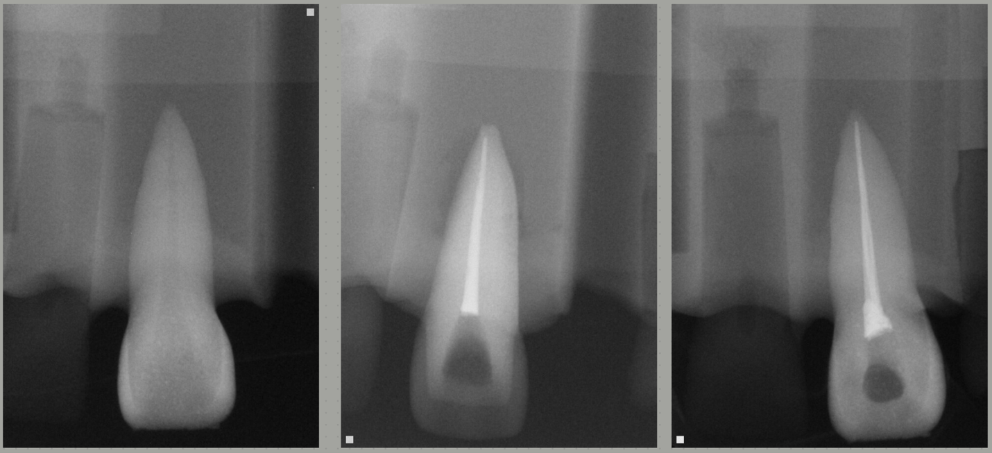
Figure (2) showing the original obturation radiograph and the substituted one
Mohamed Ibrahim, BDS, MS, DMD, PhD, MS, is Clinical Professor, Director, Pre-Doctoral Endodontics, Department of Surgical Sciences, School of Dentistry, Marquette University.
Disclaimer
The views and opinions expressed by authors are solely those of the authors and do not necessarily reflect the official policy or position of the American Association of Endodontists (AAE). Publication of these views does not imply endorsement by the AAE.