Case Challenge (Part 1)
By Dr. Bianca Aboubakare

A 20-year-old male presented for an evaluation of teeth #23 and #24. He states that he has a bump on his gums and that he had broken the teeth by trying to open a bottle six months prior and that root canal therapy was done by his general dentist soon after. The patient denies any pain with the teeth.
Medical history: Non-contributory, ASA I
Medications: None
Allergies: NKDA
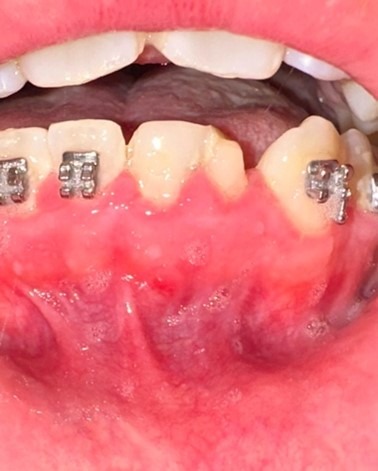
History of Present Illness: The initial treatment on #23 and #24 was done in a single visit by a general dentist 6 months prior and the teeth subsequently restored with fiber posts and resin core build-ups, and prepared for crowns. The patient had been under the care of an orthodontist for a year and presented with orthodontic brackets and wires. The patient reports that a bump appeared under his treated teeth a month prior to being seen for an endodontic evaluation.
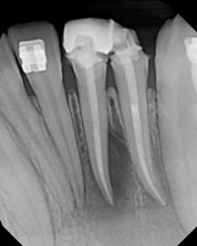
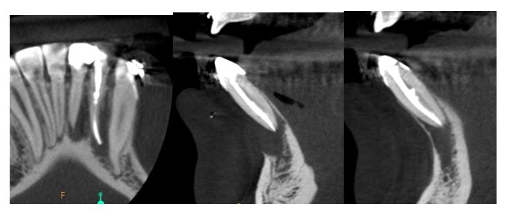
Radiographic assessment: Bone height and trabeculation appears normal with the exception of #23 and #24, which both have loss of lamina dura and periapical radiolucencies. Both #23 and #24 have coronal radiopacities consistent with temporary crowns and intracoronal radiopacities consistent with resin filling material. Both teeth have intraradicular radiopacities consistent with root filling material of adequate length, taper and density.
A limited FOV CBCT was exposed and evaluated. There are two separate well-defined areas of low density associated with the apices of #23 and #24. The lesion of #23 does not expand nor perforate the cortical plates. There is complete loss of the cortical plate of #24 and the lesion extends to the mid-root of #24 on the distal side of the root. Both #23 and #24 demonstrate high density filling material that terminates at the radiographic apices of the teeth, but the fillings are not centered. The lingual canals on both teeth are absent of high-density material.
Clinical assessment: Probing depths: 2-3 mm. #23 presents with Class I mobility and #24 presents with Class II mobility. There is a sinus tract on the buccal gingiva of #24. #23 is missing the facial aspect of its temporary crown, which is splinted with the temporary crown of #24. Both teeth are sensitive to percussion.
Diagnosis:
- Tooth #23 Previously Treated with Symptomatic Apical Periodontitis
- Tooth #24: Previously Treated with Chronic Apical Abscess
Dr. Bianca Aboubakare is an endodontist in Phoenix, Ariz., and member of the Resident and New Practitioner Committee. She completed dental school at Western University of Health Sciences, a GPR at the Veterans Affairs Medical Center in West Los Angeles, and her endodontic training at the University of the Pacific in San Francisco.