
View the winning collaborations below:
Dr. Joel Bervell “Medical Mythbuster” (Resident/Medical
Educator)
IG Reel: https://www.instagram.com/p/DNPG7yNPeLs/
TikTok:
https://www.tiktok.com/@joelbervell/video/7537499689210498318?lang=en
Dr. Adam Goodcoff (Emergency Physician)
IG Reel: https://www.instagram.com/p/DMLf-90uzjn/
TikTok:
https://www.tiktok.com/@seethemedlife/video/7527758720202329374?lang=en
Dr. Krupa Playforth (Pediatrician)
Reel: https://www.instagram.com/reel/DNYzUonBUOC/
Dr. Wendy Hunter (Pediatrician)
Reel #1: https://www.instagram.com/reel/DMtA_x_y8Lh/
About the Golden Trumpet Awards
For more than six decades, the Golden Trumpet Awards have recognized exceptional achievement in public relations, marketing, communications, and related fields across the Midwest.
The ceremony celebrates both the work shaping today’s communications landscape and the people building the future of the profession.
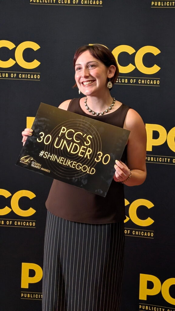 On Tuesday, Oct. 6, AAE communications specialist Rae Burach was honored as one of Publicity Club of Chicago’s 30 Under 30 at the annual Golden Trumpet Awards ceremony. The 30 Under 30 program is an annual honor recognizing rising stars and emerging leaders in public relations, marketing, communications, and media across the Chicago area. Burach joined the AAE staff in May of 2023 and has wowed colleagues and members alike ever since as a creative thinker, a natural connector and an all-around bright presence. Congratulations, Rae!
By Cami Ferris-Wong, DDS
Some endodontists say that they would like to do more restorations after their root canal treatment but they just don’t think that their referrals would allow it. Others say that they don’t feel comfortable placing restorations because they haven’t done it since dental school, or since they were in general practice. I understand both of these concerns, and I was in the same boat 20 plus years ago, so I’d like to share the story of how I got into the practice of almost every tooth leaving my office with a permanent restoration in place.
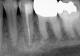
Figure 1: Example of immediate post and core restoration.
In a referral-based market, relationships are key, and my practice back in 2000 was no different. The restorative dentists wanted to restore everything, and a temporary filling was the culmination of almost every treatment I completed. I had a couple of referrals who were in insurance plans that did not pay for a core and a crown at the same visit, so they would ask me to place the core. Amalgam and core paste were the two options I had on hand and I used the former if isolation was an issue and the latter if it was a simple “fill the hole” situation. But the overall feeling was that you did what the referral asked you to do, period.
A few years later, a cascade of events happened that created a paradigm shift. First, Rick Schwartz and Ron Fransman published a beautiful article entitled “Adhesive dentistry and endodontics: materials, clinical strategies and procedures for restoration of access cavities.” 1 They outlined the scientific evidence that advocates the placement of a final restoration at the time of RCT and then went on to show how it can be done at a high level. The glossy photos of esthetically restored PFM crowns were impressive.
Around the same time, I had a patient that came in a week after I completed her RCT, complaining of a gray spot on her tooth. Her crown had been brand new when I accessed it, a standard core was placed and she was left with a less than ideal situation esthetically. She said “I just paid for a new crown and now after paying more money for the root canal, the crown looks bad.” I initially was going to tell her that there was no solution to her complaint other than a new crown, but the article in the JOE was rattling around in my brain, so I offered to redo the core procedure free of charge. This first esthetic endodontic access restoration probably took me 30 minutes to do, but it looked so much nicer than what she had and she was thrilled.
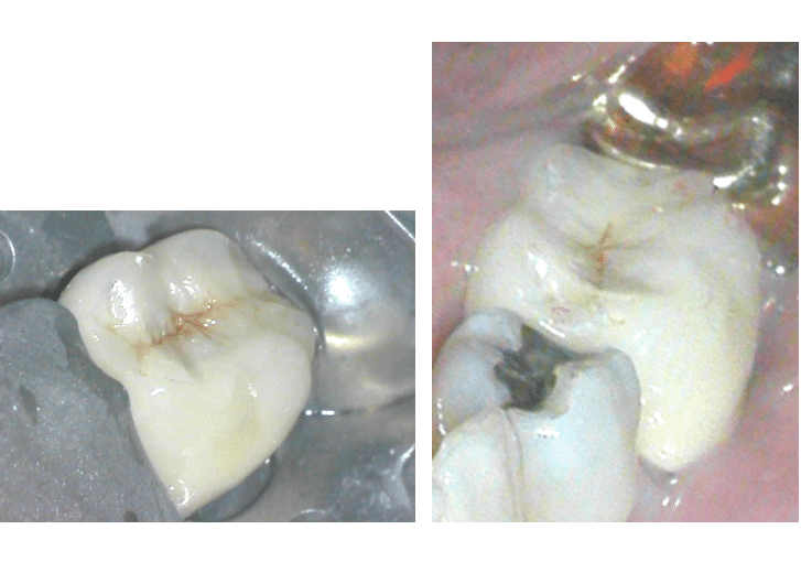
Figures 2 and 3: Endodontic access cavity permanent restoration in PFM crown filled with bulk fill composite, porcelain opaquer, macrofill composite surface, brown and white esthetic tints.
The technique for masking the gray shadow of the metal under the porcelain was detailed and time-consuming, requiring opaquer, tints, paint brushes and more. But with practice I was able to create an esthetic result and get the technique down to about 15 minutes. Fortunately, Zirconium crowns are much easier to restore esthetically, which is now a more common restoration in my practice.
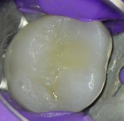
Figure 4: Zirconium crown permanent access cavity restoration.
Additionally, I was tired of seeing patients come back in with issues because they never had the tooth permanently restored. Either they came in with a fractured tooth because they bit on something hard, or the filling had fallen out and the access had been bathing in saliva. It was a frustrating scenario. I had a relationship with a local low-income clinic at the time who was booked out for months. I spoke with the director of the clinic and explained that I was concerned about the teeth not being restored right away, offering to do the cores and post and cores at a discount for their patients. She agreed that it would lessen a burden on them and that it would in fact benefit their patients.
Meanwhile, one of my best referrals and I decided to start a study club. In order to do this, we went to lunch almost every month together. Recognizing her restorative prowess, I picked her brain about the materials she used, techniques, and best practices and incorporated those things into my practice. She saw how dedicated I was to doing high-level restorative work and understood my concerns about rubber dam isolation and contamination issues. She started asking me to do the restorative on her cases and we would continue to swap tips and tricks over the years.
As for the rest of my referral population…change did not come overnight. I started slowly, calling the referral doctor to ask for permission to do the final restoration in certain cases. The main reasons that I asked to do the restorative were:
- The patient is very anxious and we were doing treatment under oral or I.V sedation. These were usually a slam dunk, because the referring doctor doesn’t typically want to deal with an anxious patient any more than we do. (Kids fall into this category as well).
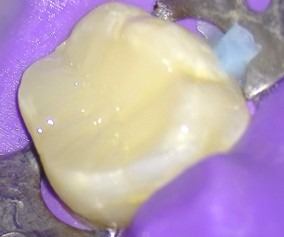
Figure 5: Build-up on tooth #19 for a 13 year-old boy.
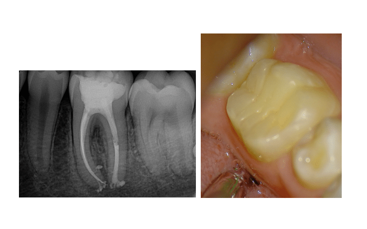
Figure 6 and 7: 1 year recall radiograph and photograph of tooth #19 on the same patient
2. The patient is leaving to go out of town or requests the final restoration. This happened often because I would explain the treatment and that they have to go back to their restorative dentist to fill the access cavity and they would say “Can’t you just do that?”
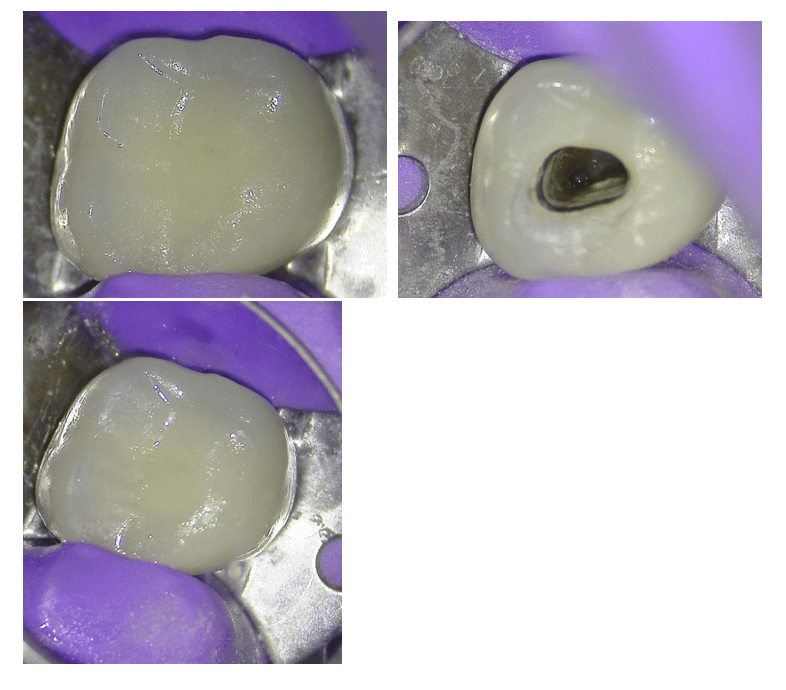
Figures 8-10: Pre-op photo, picture taken during treatment and post-op photo of tooth #2 PFM access restoration.
3. For teeth that have recurrent caries, deep decay is a good reason to ask to do the restoration (once you feel comfortable doing it). The general dentist is likely not going to place a rubber dam and we know that a post and core restoration will fare better with proper isolation 2. The dentist is generally thrilled if you are willing to remove the crown and caries and then restore the tooth afterward, making their crown prep much easier. (In these cases we take a pre-op impression and send it to the referral in case it helps them in planning or making the temporary crown.)
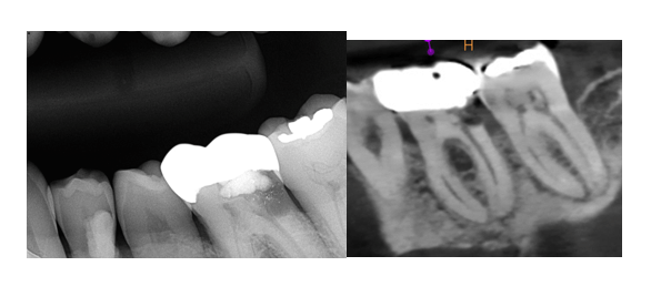
Figures 11 and 12: Pre-op radiograph and sagittal CBCT slice of tooth #19.
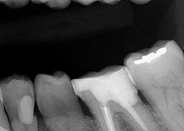
Figure 13: Post-op bitewing of post and core restoration.
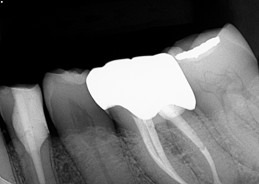
Figure 14: Bitewing radiograph after crown placement.
Over the years, my referrals began to understand how my completing the final restoration could benefit their practice, allowing them to do high-value restorative procedures instead of spending time on the build-up or an access cavity restoration. Additionally, patients are happy to leave with something solid in their mouth that will not break down over time. It is a worthwhile, albeit often gradual, endeavor that takes vision, strategy and a commitment to excellence.
References:
- Schwartz, R.S. ∙ Fransman, R. Adhesive dentistry and endodontics: materials, clinical strategies and procedures for restoration of access cavities: a review. J Endod. 2005; 31:151-165
- Goldfein J, Speirs C, Finkelman M. Amato R. Rubber dam use during post placement influences the success of root canal–treated teeth.
J Endod, 2013; 39, 1481-1484
Disclaimer
The views and opinions expressed by authors are solely those of the authors and do not necessarily reflect the official policy or position of the American Association of Endodontists (AAE). Publication of these views does not imply endorsement by the AAE.
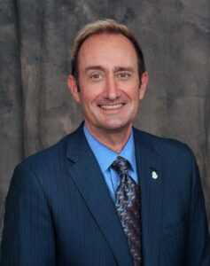 It’s that time of year….School is back in session. The leaves are changing color, the crisp Autumn air is beginning to set in. So now, all of our thoughts turn to: Football? Maybe – but that would depend on how well your team is performing. The holidays? Maybe later. It’s just too early. But the one big thing on every Endodontist’s mind is, of course, Insight Track! Our annual Fall meeting is extremely popular and provides a smaller, more intimate CE experience compared to our larger annual meeting in the Spring. This year we will be meeting in beautiful Puerto Rico at the Hyatt Regency Grand Reserve just outside of San Juan. This venue will provide an idyllic tropical getaway to prepare you for the winter months ahead. But more importantly, our educational program will inspire you and undoubtedly reinforce your enthusiasm for our specialty of Endodontics. This year’s topic is Trauma – a timely subject, coming on the heels of our recently published update of our Trauma Guidelines.
 Compiled by Elisabeth Lisican
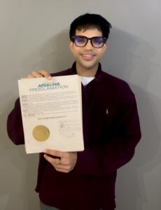
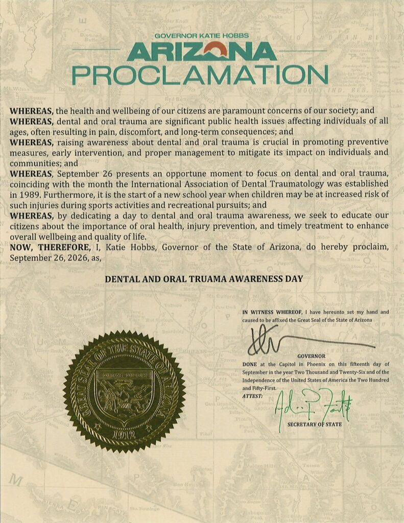
Ahead of Global Dental & Oral Trauma Day on Sept. 21, one Arizona dental student took an extra step to put dental trauma education in the spotlight. Drawing on a personal experience with dental trauma that helped inspire a future career in dentistry, the student successfully pursued an official proclamation from the state of Arizona recognizing the day.
In the Q&A below, Samved Nambiar shares the personal motivation behind the effort, the steps involved in securing the proclamation and how mentors within the dental trauma community helped along the way. The conversation also explores an early interest in endodontics and plans to use the proclamation as a springboard for greater awareness among fellow students, parents and the broader community.
Elisabeth Lisican: Why did you decide to pursue a proclamation to mark this very special date for dental and oral trauma awareness?
Samved Nambiar: I wanted to pursue a proclamation for dental trauma because this is an issue that resonates deeply with me. I experienced dental trauma as a child, and my journey with a broken front tooth and trying to restore my smile is actually what planted the dream of becoming a dentist in me in the first place.
I still remember feeling helpless and not knowing what I should do. Would my tooth grow back? What were my options? What would happen next? Those are questions that I think many children and parents can have when dental trauma happens.
With this proclamation, I hope to help spread awareness, especially among children and their parents, that dental trauma is real, but with the right information and timely action, it does not have to become a long or frightening journey. I hope to use this opportunity to share resources and promote awareness about what people can do when dental trauma occurs.
Lisican: What steps did you have to take to secure the proclamation?
Nambiar: The first person I contacted was Dr. Katie Divine, who I consider one of my mentors and who is actually the reason I joined IADT. She helped me understand how a proclamation should be approached and gave me an idea of the language and formal requirements I needed to consider.
I also contacted the Arizona Governor’s Office, and they explained the process, deadlines, and drafting requirements necessary for a proclamation to be considered. Even after learning the process, it still felt like a pretty big task.
I then reached out to Dr. Bradley Gettleman, whose lectures at ASU I have attended regularly. I knew he had previously helped secure a proclamation in Arizona, so I asked for his guidance as well. His experience helped me understand how to move forward.
Once I had a better understanding of the process, I drafted the proclamation and submitted it to the Governor’s Office. From there, I stayed in regular communication with their office and made sure I was available to provide anything else they needed throughout the process.
Lisican: How did you first get interested in the IADT?
Nambiar: I first learned about IADT at a pre-dental lecture where Dr. Katie Divine was a guest speaker. She introduced me to the organization and its work, and I immediately became interested. I ended up paying my IADT student membership dues and joining soon after.
IADT resonated with me because dental trauma is something I have personally experienced. In many ways, it is part of the reason I became interested in dentistry in the first place.
Beyond my personal connection, I also became interested in the broader importance of dental trauma education. When an injury happens, especially to a child, parents and caregivers may not know what to do or how urgently they need to act. Being part of IADT gives me an opportunity to learn more about dental trauma while also helping spread awareness and making sure families feel better equipped to respond when it happens.
Lisican: What got you interested in dentistry?
Nambiar: I have always known that I wanted to pursue healthcare. My dad is a physician, and my brother is an epidemiologist, so growing up, I thought I would follow the pre-med route.
My love for dentistry really began with the endodontist who treated the front tooth I had broken as a child. Before that, I had experienced dental anxiety and did not always have great experiences at the dentist. I remember being told that getting a root canal would hurt, so I was extremely nervous going into the appointment.
But the procedure was completely different from what I expected. The doctor walked me through every step, explained what he was doing, and made me feel comfortable throughout the process. I remember leaving that appointment thinking that I wanted to become the kind of doctor who could help patients feel that same sense of trust and comfort.
After that, I started shadowing different areas of dentistry, including general dentistry, endodontics, periodontics, and oral surgery, and I kept falling in love with the profession more.
I love the hands-on aspect because I have always enjoyed building things and working with my hands. I also really enjoy the leadership and management side of dentistry, working with a team, making sure people feel heard, and creating an environment where everyone can work efficiently together.
For me, dentistry brings together healthcare, hands-on work, patient relationships, and leadership in a way that feels very natural to me.
Lisican: What are your plans for the special month of September?
Nambiar: On September 26, I will be hosting a meeting with members of the ASU Pre-Dental Society. I currently have the privilege of serving on the executive board, and I want to use that platform to introduce more students to dental and oral trauma awareness and the resources available through IADT.
I was also able to work with IADT to obtain a discount code for dental trauma awareness merchandise, which I plan to share with students as another way to help spread the message.
Beyond that, I am planning an Instagram Reel campaign to help bring awareness to dental trauma and encourage people to learn more about prevention and what to do when an injury occurs.
For me, the proclamation is really the beginning. I want to use the recognition as a platform to keep the conversation going and reach more students, parents, and members of the community.
Elisabeth Lisican is the AAE’s assistant director of communications & publishing.
APICES 2026 brought endodontic residents from across the country to St. Louis for two days of learning, conversation and connection beyond the residency experience. The program tackled the questions residents are thinking about as they prepare for the next stage of their careers. Dr. Gordon Lai shared lessons in Things I Wish I Knew in Endo Residency, while Dr. Monica Estes explored compensation in today’s market. Dr. Rene Chu offered perspective on postgraduate life, Dr. Rob Roda took on the importance of referral relationships in 10 Ways to Lose a Referral, and Dr. Adham Abdel Azim closed the educational program with a clinical look at cervical root resorption. Residents also had the opportunity to learn more about the profession through Get to Know Your Specialty, featuring Drs. Joseph Dutner, Craig Hirschberg, W. Craig Noblett and Yehuda Joseph Benjamin. Between sessions, the exhibit hall, meals with corporate partners and social events at Ballpark Village gave residents time to connect with colleagues from programs across the country and with the broader endodontic community. Together, the weekend offered a mix of practical career insight, clinical education and the kind of peer connection that makes APICES a unique part of the resident experience.
Click here to view photos from APICES and Educator Workshop!
Save the Date: APICES 2027
August 20–21, 2027
Marriott New Orleans | New Orleans, Louisiana
Registration is expected to open in early July 2027, with additional program details to come.
The 2026 Educator Workshop, held August 14–15 in St. Louis, brought together endodontic program directors from across the country for two days of education, candid discussion and collaboration focused on the challenges and opportunities facing advanced endodontic education. The program covered a wide range of timely topics. The American Board of Endodontics shared updates on a record 166 new Diplomates, its completed Job Task Analysis, updated examination domains and changes to the examination process. Sessions also explored AI in education, CODA updates, ADEA PASS and the Dental Match, giving attendees an opportunity to better understand changes affecting both educators and residents.
A major focus of the workshop was the future of the educator workforce. Program directors discussed the challenges of recruiting and retaining faculty, noting that barriers to academic careers extend beyond compensation. Early identification of future educators, mentorship, part-time teaching opportunities and stronger pathways into academics were among the strategies discussed. Attendees also spent significant time examining the residency application, interview and offer process, including opportunities for greater consistency and clearer expectations across programs.
Just as valuable as the formal presentations were the conversations among program directors themselves. The workshop provided dedicated time to compare experiences, discuss common challenges and identify areas where continued collaboration could support both educators and residents. As a next step, the AAE Educational Affairs Committee will develop a resident survey focused on interview timing and the residency application experience, providing additional insight to inform future discussions. More information on the survey will be shared as that work progresses. These conversations and next steps will continue to inform the work of the Educational Affairs Committee in the year ahead.
Check out our Flickr album of photos from Educator Workshop and APICES!
Save the Date: Educator Workshop 2027
The next Educator Workshop will take place August 20–21, 2027, at the Marriott New Orleans in New Orleans, Louisiana, and will focus on predoctoral directors. The 2027 program will include two joint presentations with Corporate Community Conference attendees, along with dedicated programming developed around the priorities and challenges identified by predoctoral educators. Attendees from both groups will also have an opportunity to connect at a joint Educator and Corporate Community Welcome Reception on Thursday, August 19, before the program begins. Registration is expected to open in June 2027. Additional program and registration information will be shared with predoctoral directors as planning continues.
By Ane Poly, DDS, MSc, PhD
As we navigate the rapidly evolving landscape of dental education, exploring how digital technologies can enhance current teaching methods and improve student learning is paramount. Endodontic access preparation (EAP) represents the critical first phase in non-surgical root canal treatment. Its primary objectives, such as conserving sound tooth structure, unroofing the pulp chamber, and establishing direct-line access to canal orifices, lay the foundation for clinical success.
Traditionally, preclinical courses rely on visual inspection by calibrated faculty members to grade student EAP. However, extensive literature shows that visual inspection is inherently subjective, leading to a lack of interrater reliability among faculty in the evaluation process.
Standard visual inspection also leaves students with limited, qualitative feedback. While an instructor can observe that an access cavity is overextended or off-center, students often struggle to visualize the exact three-dimensional volumetric defect or millimeter deviations from ideal dimensions. Because self-assessment is a crucial lifelong skill predoctoral students must develop to refine their hand-skills and work independently, establishing objective standards for error detection is essential.
Adapting 3D Comparison Software for Endodontics
To bridge this gap, CAD/CAM technology widely used in fixed prosthodontics to evaluate crown preparations (Planmeca Romexis Compare, formerly E4D Compare) was adapted for endodontic education to evaluate student EAPs three-dimensionally against standardized ideal master models.1
Students start by using intraoral scanners to scan their EAPs and upload the 3D file into the software. Then, they trace the access perimeter as an “Axial Base” and follow the board licensure landmarks (ADEX criteria2) to trace the “Margin”. High-resolution surface scanning superimposes the student model onto a faculty-determined ideal model, and the existing software tools are used to measure internal wall dimensions and outline extents:
- Compute Difference: Quantifies volumetric alignment via 3D heat maps, highlighting overextended areas in red, underextended areas in blue, and acceptable areas in yellow. (Figure 1)
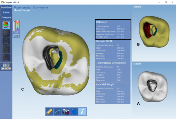
Figure 1: Compute difference tool showing (A) the ideal model, (B) the S2 model, and (C) the two models overlapped. Red areas indicate overextension; blue areas indicate underextension; yellow areas fall within the tolerance range. The black rectangle highlights the quantitative data.
- Slice Plane & Distance Tools: Provide cross-sectional views at various depth levels, enabling specific millimeter measurements between internal preparation walls. (Figure 2)
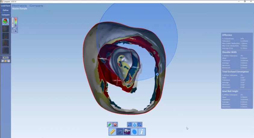
Figure 2: Ideal and student models overlapped with the slice plane tool showing a qualitative comparison of the internal form. Three measurements are shown in specific areas, illustrating the distance tool being used to measure the distance between models.
- Shoulder Width: Measures circumferential boundary distances to verify adherence to licensure safety margins.2 (Figure 3)
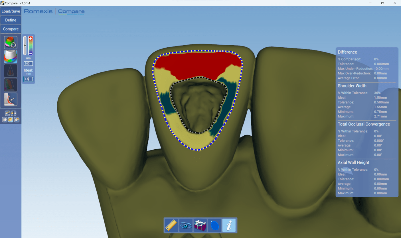
Figure 3: Shoulder width tool shows the student model where the red areas indicate the distance is > 2.0 mm, blue areas < 1.0 mm, and yellow areas between 1 and 2 mm.
Practical Insights from Preclinical Implementation
The viability and impact of this digital methodology were evaluated with a randomized controlled trial.3 Sixty second-year dental students performed EAPs on #14 acrylic teeth (RTE #14 With Insert; Acadental, Lenexa, KS) following the ADEX criteria2. Students were assigned either to a control group using traditional visual assessment or an experimental group combining traditional self-assessment with 3D software evaluation.
The trial revealed significant findings regarding student self-perception and critical evaluation. While both groups experienced an increase in clinical confidence, students relying solely on traditional visual assessment significantly overestimated their performance improvements. Conversely, students equipped with 3D digital feedback examined their work far more critically and accurately.
This outcome highlights the Dunning-Kruger effect, a cognitive bias where individuals with lower competence overestimate their abilities due to a lack of detailed feedback. The 3D comparison software serves as a “digital magnifying glass,” exposing minor over-extensions, wall gouging, and lack of straight-line access. By removing visual ambiguity, the software tempers unearned overconfidence and fosters genuine self-awareness.
Looking Ahead: Impact on Dental Curriculum and Faculty Workflow
Student acceptance has been positive. In our randomized trial, 96.7% of students who used the 3D method agreed that 3D evaluation should be permanently incorporated into preclinical endodontic training and licensure exam preparation.
Beyond student self-evaluation, integrating 3D evaluation software into preclinical simulation laboratory training helps address a broader institutional challenge: faculty shortages and calibration. With vacant budgeted faculty positions in U.S. dental schools reaching record highs, digital evaluation provides an objective, standardized baseline for instruction.
Ultimately, this precise and immediate feedback empowers students to critically analyze their work and build the lifelong self-assessment capabilities necessary for modern endodontic practice. Furthermore, by providing round-the-clock feedback, the software mitigates faculty workload, allowing instructors to redirect their time toward targeted, high-level clinical coaching.
Note: Generative AI was used as an editorial aid to assist with grammar, clarity, phrasing, and organization. The ideas, opinions, and final wording of the content are solely those of the author.
Ane Poly, DDS, MSc, PhD, is a Clinical Assistant Professor in the Department of Endodontics at the University of Florida College of Dentistry.
References
- Poly A, Burnett JE, Buie CA, Schweitzer JL. Three-dimensional software adapted to evaluate endodontic access cavity preparation. J Dent Educ. 2023;87(suppl 3):1848-1851.
- CDCA-WREB-CITA. ADEX Dental Exam Series: Endodontic Criteria. 2026 Candidate Manual. Anterior Endo Procedure: https://adextesting.org/wp-content/uploads/2026/05/2026.8-DEN-ENDO-Anterior_20260521.pdf Posterior Endo Procedure: https://adextesting.org/wp-content/uploads/2026/05/2026.8-DEN-ENDO-Posterior_20260521.pdf
- Poly A, Harness C, Vu E, Biradar A, Buie CA, Burnett JE, Schweitzer JL. Integrating digital technology in endodontic education: A randomized controlled trial evaluating student self-assessment and perspectives. J Dent Educ. 2025 Aug;89(8):1294-1302.
 Compiled by Dr. Blake Clarke
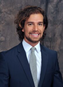
Dr. Jan Groth is a resident at the University of Minnesota. Here, Dr. Blake Clarke catches up with him and learns more about his journey to endo.
Dr. Blake Clarke: Dr. Groth, thank you for taking the time to speak with us. Could you begin by telling us a little about yourself and your background?
Dr. Jan Groth: Thanks for having me! I’m an endodontics resident at the University of Minnesota.
Before returning for residency, I spent several years in private practice as a general dentist in Minneapolis and Miami Beach. Over time, I noticed that the days I went home feeling most fulfilled were the ones when someone arrived miserable from a severe toothache and left comfortable, often surprised that the root canal was not the horror story they had imagined. I genuinely enjoy the psychological side of what we do: caring for someone who is terrified by the words ‘root canal,’ earning that patient’s trust, keeping them comfortable, and turning the experience into a positive one.
Outside dentistry, I enjoy staying active and exploring. I love snowboarding and taking on major treks through different landscapes, whether on high-altitude trails or in remote terrain. I also enjoy deep conversations, learning about space, caring for plants, and working with my hands on design and woodworking projects. I believe in making time to enjoy life and finding meaningful ways to give back to the community around me.
Dr. Clarke: You practiced as a general dentist for several years after graduation. When did you decide to pursue endodontics, and what led you to that decision?
Dr. Groth: Practicing as a general dentist gave me the opportunity to explore a broad scope of care while navigating different practice cultures, patient communities, and procedures. Although I have always loved dentistry, consecutive years of undergraduate education, research, and dental school had absorbed a significant chapter of my life. After graduation, I felt it was important to step away from academia, gain real-world perspective, and reconnect with life and the community around me. I always had an eye on endodontics, but working as a general dentist first gave me invaluable clinical perspective and personal balance.
About four years into practice, I reached a transitional point in my life and knew I would be moving. That gave me space to step back and reevaluate what truly drove me. I realized I wanted deeper knowledge and the opportunity to refine my skills in the areas where I felt the greatest passion and natural strengths: saving teeth and helping patients manage anxiety. With that clarity, returning to specialize felt like the right decision.
Dr. Clarke: You mentioned that finding meaningful ways to give back is important to you. How do you hope to carry that into your career after residency?
Dr. Groth: Growing up with Mexican and German heritage, connecting across different cultures has always felt natural to me. Serving on dental brigades abroad, including in Guatemala, showed me how powerful those connections can be. Using my native Spanish to communicate directly with patients allowed me to address some of the fear that often surrounds dental care.
Long term, I hope to establish a sustainable brigade in Latin America that provides free specialty dental care while partnering with the local community to support its growth beyond the dental chair.
Many patients arrive for endodontic treatment already anxious about the procedure. What have you learned about earning their trust and helping them feel more comfortable?
Most patients don’t come in with only a painful tooth. They also bring years of accumulated dental anxiety and secondhand horror stories, all while being asked to put themselves in a vulnerable position. I’ve learned that earning their trust begins with genuine empathy.
Before I recline the chair or pick up a mirror, I make a point of connecting with them on a human level. I meet them eye-to-eye, validate their apprehension, and make sure they retain a sense of control.
When patients realize that you see them first and their root canal second, their anxiety often begins to ease. Turning an appointment someone has dreaded for weeks into a comfortable, almost anticlimactic part of the day is one of the most rewarding aspects of this specialty.
Dr. Clarke: That is an excellent approach to caring for patients with dental anxiety. For our final question, what is one piece of advice you would give to dental students or general dentists who are considering a career in endodontics?
Dr. Groth: My advice would be to give yourself grace and move with intention rather than simply following momentum. Whether you are accepted directly out of dental school or spend valuable years in general practice, both paths have real advantages. What matters most is giving yourself the space to step back and reconnect with what truly motivates you. It is easy to get caught up in academic milestones, but when you pursue endodontics out of a genuine passion for saving teeth and relieving pain, the demands of the profession feel worthwhile.
Dr. Blake Clarke is a member of the AAE’s Resident & New Practitioner Committee.
By: Dr. Jessica Hwang
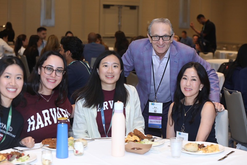
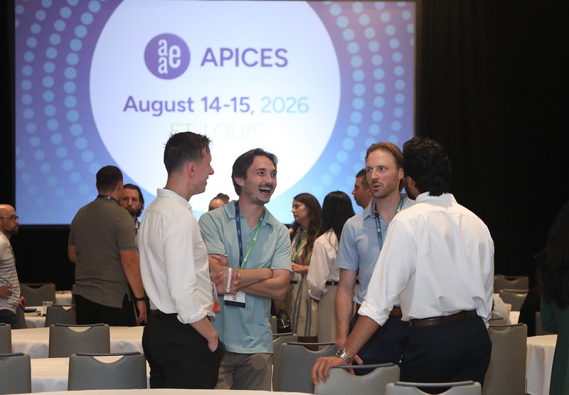
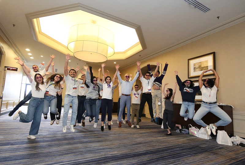
This August, endodontic residents from across the country came together in St. Louis for the annual APICES conference, and the weekend was a great reminder that some of the most valuable parts of residency happen outside of the clinic.
Held August 14–15 at the Hyatt Regency St. Louis at the Arch, APICES brought residents together for two days of education, career development, networking, and plenty of opportunities to have some fun along the way. The program covered many of the questions residents are already thinking about as they prepare for life after training. Dr. Gordon Lai kicked things off with Things I Wish I Knew in Residency, sharing invaluable advice ranging from patient management and optimizing resident workflows to the practical clinical tips he is well known for.
Another crowd favorite was Dr. Monica Estes’ presentation, Beyond the Percentage: Understanding Endodontic Compensation in Today’s Market. Dr. Estes walked residents through real-world compensation scenarios, common contract considerations and potential red flags, and how to more thoughtfully evaluate employment opportunities. Residents also heard from Dr. Rene Chu on life after residency, a topic everyone in training is eager to think about! On the clinical side, Dr. Adham Azim delivered an engaging surgical lecture on the diagnosis and management of cervical root resorption, while Dr. Rob Roda shared plenty of wisdom and a few cautionary lessons in 10 Ways to Lose a Referral. Residents also heard from leadership across the AAE, AAE Foundation, and ABE College of Diplomates during the annual Get to Know Your Specialty session.
Beyond the lecture hall, the exhibit hall stayed busy throughout the weekend, with industry partners showcasing new technology, answering questions, and keeping things lively with giveaways and raffles. It gave residents an opportunity not only to learn about products and resources they may encounter in practice, but also to connect directly with the companies and organizations supporting the specialty.
APICES also provided an opportunity to give back. Before the meeting began, members of the Foundation Resident Expert Advisory Council (REACH) volunteered with Give Kids a Smile St. Louis, helping prepare dental supplies for the organization’s biannual open clinic in October. It was a meaningful way for residents to contribute to the local community while representing the Foundation for Endodontics and the specialty.
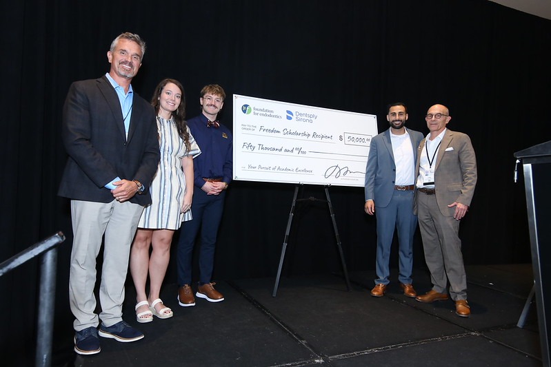
One of the weekend’s special moments was recognizing the 2026 recipients of the Foundation for Endodontics and Dentsply Sirona Freedom Scholarship. Each year, three residents receive a $50,000 award designed to help offset the significant financial investment of postgraduate endodontic training. This year’s recipients were Dr. Luc de la Villefromoy, Dr. Anas Akhras, and Dr. Sierra Nunn. The scholarship is another meaningful example of the support being invested in the next generation of endodontists.
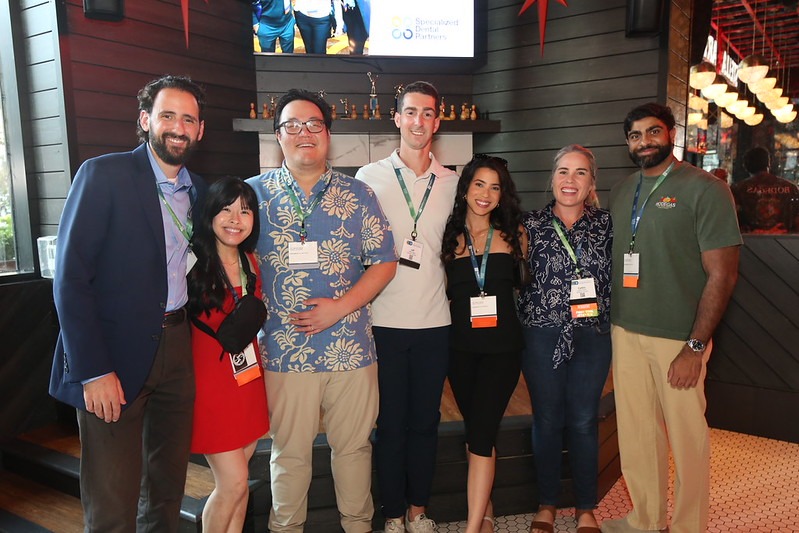
Of course, APICES would not be APICES without the social side of the weekend. Corporate partners Specialized Dental Partners and Specialty1 Partners hosted events throughout St. Louis, giving residents a chance to step away from lectures, enjoy stunning views of the Gateway Arch, and connect in a more relaxed setting. Between the receptions, exhibit hall conversations, and catching up with residents from programs across the country, there was no shortage of opportunities to make new connections.
That may ultimately be what makes APICES so memorable. Residency can be intense, and it is easy to become absorbed in the day-to-day routine of patients, seminars, research, and requirements. APICES gives residents a chance to look beyond their own programs and realize just how large and welcoming the endodontic community really is. Hopefully, residents walked away with new knowledge, new friends, and a stronger sense that they have found their place in the specialty.
Until next year, APICES!
Dr. Jessica Hwang is a member of the AAE’s Resident & New Practitioner Committee.