Genes at the Root: Unlocking the Genetic Mechanisms of Apical Periodontitis
By Ariadne Letra, DDS, MS, PhD
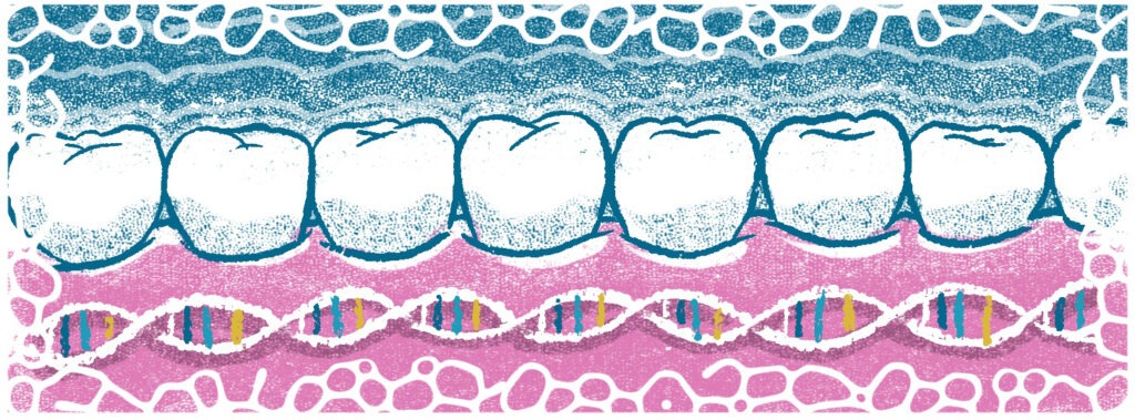
Apical periodontitis (AP) results from the progression of microorganisms within an infected/necrotic root canal system. The localized infection activates the local host immunoinflammatory response, triggering a cascade of events with recruitment of immune cells, release of inflammatory mediators, establishment of local inflammation, hard tissue breakdown, and eventual formation of a periapical lesion1.
Studies in humans and animal models have implicated host factors as critical contributors to AP susceptibility, with host-pathogen interactions suggested to influence disease development and progression1. Host factors involving genetic regulatory mechanisms may affect host physiology, resulting in imbalances in the expression of key pro- and/or anti-inflammatory mediators that integrate the complex biological network underlying AP.
In other words, when an individual’s genetic machinery is disrupted, the consequences may affect how the immune system responds to infection, whether a patient heals, how much pain they experience, and even whether they develop the disease at all2.
Genetic mechanisms such as DNA variation (e.g., single-nucleotide polymorphisms, SNPs) have been proposed as host factors potentially influencing an individual’s risk of AP development, progression, and/or repair. SNPs are the simplest form of genetic variation among people and occur on average about once in every 1,000 nucleotides, which means that each person has roughly 4 to 5 million SNPs in their genome. Most SNPs are neutral, but those located within functionally important genes can alter gene and protein expression, impair cellular function, and thereby influence disease susceptibility or healing capacity2,3.
What the Research Shows
In recent years, emerging evidence has supported AP as a genetically regulated process with overlapping protective and destructive functions. Studies across multiple populations have found that polymorphisms in disease-relevant genes, including pro-inflammatory mediators such as interleukins, matrix metalloproteinases, tumor necrosis factor, heat shock proteins, and others, are associated with AP. The majority of these studies reflect candidate genes selected based on their reported function in the pathogenesis of AP, and revealed the association of many genes, such as IL1B, IL6, IL8, MMP1, MMP3, MMP8, TNFA, TBX21, HSPA1L, WNT3A, among others 2-6. One variant in IL1B (rs1143643) that was significantly associated with AP was also shown to alter gene and protein expression in periapical tissues5. Similarly, the MMP1 promoter variant (-1607 1G/2G) associated with AP was also shown to result in differential gene expression based on individual genotypes; individuals with this polymorphism had increased MMP1 mRNA expression in periapical tissues as compared to healthy tissues obtained from individuals without AP4. Among the more clinically promising findings is the role of the WNT signaling pathway in bone repair. WNT3A variants are associated with AP, and laboratory evidence suggests WNT3A may be a viable therapeutic target for accelerating bone healing after AP-related bone loss6,7, pointing towards targeted treatment possibilities. These findings suggest functional effects of these polymorphisms on cellular functions involved in AP pathogenesis and shed light on how polymorphisms may shape an individual’s risk of AP and how they may be used as targets for treatment strategies.
Genome-Wide Studies
Recently, two genome-wide association studies analyzing over two million genetic variants in thousands of adults with and without AP revealed the association of novel genes with AP, with and without associated pain8,9. Among the newly associated genes are RAP1GAP (RAP1 GTPase activating protein) and SPP1 (osteopontin), both of which are involved in immune cell recruitment, macrophage polarization, and regulation of immune-inflammatory response8. Moreover, these studies found distinct male-only and female-only AP-associated variants, confirming a sexual dimorphism pattern reported in epidemiological studies and animal models of AP, highlighting potential implications for precision AP risk assessment and treatment planning10.
Toward Precision Endodontics
A deeper understanding of the genetic underpinnings in AP is essential for developing precision diagnostic and treatment strategies. That said, the associations identified to date should not be interpreted as causation. Genetic association studies remain limited by sample size, population diversity, and difficulty controlling for environmental confounders such as microbiome variation. Therefore, the most significant advances in the field will be made through unbiased studies with large and diverse populations and functional validation of identified SNPs as potential targets for precision treatment strategies8.
For now, endodontic treatment remains the standard of care, and most patients heal well. But the trajectory of this research points toward a future in which genetic profiling may help clinicians predict a patient’s risk of developing AP, likelihood of treatment success or failure, susceptibility to associated pain, and optimal pharmacological management, enabling more predictable diagnosis, treatment, and prognosis of all endodontic patients. While such precision endodontic approaches are not yet a clinical reality, recent research advances suggest they are within reach.
References
- Cavalla F, Letra A, Silva RM, Garlet GP. Determinants of Periodontal/Periapical Lesion Stability and Progression. J Dent Res. 2021:100:29-36.
- Menezes-Silva R, Khaliq S, Deeley K, Letra A, Vieira AR. Genetic Susceptibility to Periapical Disease: Conditional Contribution of MMP2 and MMP3 Genes to the Development of Periapical Lesions and Healing Response. J Endod. 2012; 38:604-607.
- Letra A, Ghaneh G, Zhao M, Ray H, Francisconi CF, Garlet GP, Silva RM. MMP -7 and TIMP-1, new targets in predicting poor wound healing in apical periodontitis. J Endod 2013; 39:1141-1146.
- Trombone AP, Cavalla F, Silveira EM, Andreo CB, Francisconi CF, Fonseca AC, Letra A, Silva RM, Garlet GP. MMP1-1607 polymorphism increases the risk for periapical lesion development through the upregulation MMP-1 expression in association with pro-inflammatory milieu elements. J Appl Oral Sci. 2016; 24:366-375.
- Dill A, Letra A, Souza LC, Yadlapati M, Garlet GP, Vieira AR, Silva RM. Analysis of multiple cytokine polymorphisms in individuals with untreated deep carious lesions reveals IL1B (rs1143643) as a susceptibility factor for periapical lesions development. J Endod. 2015; 41:197-200.
- Souza LC, Cavalla FC, Maili L, Garlet GP, Vieira AR, Silva RM, Letra A. WNT gene polymorphisms and predisposition to apical periodontitis. Sci Rep. 2019; 9:18980.
- Tang Y, Zhou X, Gao B, Xu X, Sun J, Cheng L, Zhou X, Zheng L. Modulation of Wnt/β-catenin signaling attenuates periapical bone lesions. J Dent Res 2014;93:175-182.
- Petty LE, Silva RM, Souza LC, Vieira AR, Shaw DM, Below JE, Letra A. Genome-wide Association Study Identifies Novel Risk Loci for Apical Periodontitis”. Petty LE, Silva R, de Souza LC, Vieira AR, Shaw DM, Below JE, Letra A. J Endod. 2023;49:1276-1288.
- Salminen A, Hyvärinen K, Ritari J, Leppilahti JM, Palotie U, et al. Genome-wide association study of pulpal and apical diseases. Nat Commun. 2025 Jul 23;16(1):6774.
- Sangalli L, Souza LC, Letra A, Shaddox L, Ioannidou E. Sex as a Biological Variable in Oral Diseases: Current Perspectives and Future Directions. J Dent Res. 2023, 102(13): 1395–1416.
Ariadne Letra, DDS, MS, PhD, is Professor and Assistant Dean for Faculty Affairs, Department of Oral and Craniofacial Sciences, Department of Endodontics, Center for Craniofacial and Dental Genetics, at the University of Pittsburgh. She is also an associate editor of the Journal of Endodontics. Dr. Letra can be reached at AriadneLetra@pitt.edu.
Disclaimer
The views and opinions expressed by authors are solely those of the authors and do not necessarily reflect the official policy or position of the American Association of Endodontists (AAE). Publication of these views does not imply endorsement by the AAE.