
Decision-making for Managing Immature Permanent Teeth with Necrotic Pulp
By Hacer Aksel, DDS, PhD
As clinicians, we often face complex treatment decisions in daily practice. From my experience, managing necrotic immature root canals was one of the topics residents most frequently asked about when approaching these cases. This article summarizes key considerations to support clinical decision-making.
Root development is classified into five stages according to Cvek’s classification. As a general consensus, immature permanent teeth with necrotic root canals and root development at stages 1 (less than 1/2), 2 (1/2), or 3 (2/3) can be treated with regenerative endodontic therapy (RET) to promote further root development. At stage 4 (near full-lengthening with an open apex), either RET or apexification can be performed (1). RET allows tissue formation and root maturation, whereas apexification creates a hard tissue barrier using long-term calcium hydroxide or a calcium-silicate apical plug. A review of cohort studies reported similar success and survival rates for RET and MTA apexification, supporting RET as a first-line treatment for immature permanent teeth with pulp necrosis (2). However, root maturation with RET varies and carries a higher risk of adverse postoperative events, including persistent infection, tooth discoloration, fracture, and root resorption, highlighting the importance of thorough discussions before finalizing treatment.
The single-visit apical plug technique became possible with the development of calcium silicate-based materials (CSMs). Materials that do not cause discoloration, have good handling and physical properties, are less cytotoxic, and set quickly are popular in practice. Apical barrier formation with CSM is independent of the prior use of calcium hydroxide. When exposed to tissue fluids, CSM forms hydroxyapatite and calcium hydroxide, providing bioactivity and antibacterial activity. A 4-5 mm apical plug can provide an adequate seal (3). However, extrusion through the periapical tissues can occur during placement. To minimize extrusion, fast-setting materials can be preferred. Additionally, a collagen-type apical barrier can help but may not prevent extrusion. Gentle, incremental placement of fast-setting CSM in small amounts (2-3 mm thick), along with periodic periapical radiographs, can help control placement. In cases of severe inflammation or drainage, the inflamed area may have a pH as low as 5.5. Acidic contamination can affect proper setting, strength, antibacterial properties, and hard tissue formation (4). In these cases, initial calcium hydroxide medication can protect the physical properties and the setting reaction of CSM. After placement of the apical plug, the remaining canal space can be filled with gutta-percha and sealer, composite resin, or full-length CSM. In vitro studies reported no significant differences in fracture resistance or stress distribution between RET and apical plug formation if the tooth is properly restored (5).
Several factors influence prognosis after RET. Younger patients, especially those up to 13 years of age, and teeth at earlier stages of root development have been reported to have greater root development (6, 7). Patient age has become a key factor in case selection. A recent survey found that 56.5% of clinicians perform RET in patients aged 15 or younger, while only 22.6% consider it for those over 20 (8). Most patients undergoing RET have been reported to be under 18 years old. Only a few studies have examined RET in patients aged 20 or older, and none have reported root lengthening or thickening (9). Therefore, in older patients, especially those with a calcified barrier, an apical plug with CSM may be preferable, given lower expectations for continued root development in adults (Case 1 and Case 2).
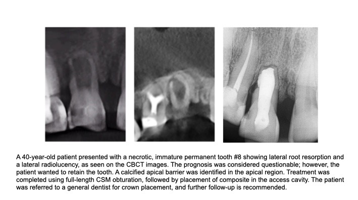
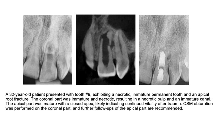
Long-term follow-up is essential for RET, making patient compliance critical. The AAE recommends clinical and radiographic follow-up at 6, 12, and 24 months, then annually after two years. Extended follow-up is important, as up to 39% of failures may not be detected until at least two years after treatment initiation (8, 10). Persistent infection is a major challenge, impairing stem cell differentiation and tissue formation, and accounts for 79% of RET failures (10). Traumatic injuries reduce RET prognosis compared to developmental anomalies such as dens invaginatus, likely due to negative effects on cellular function and vascularity (11). If RET fails, treatment decisions should consider root maturity, restorability, and the cause of failure. If the tooth is asymptomatic and the periapical lesion is healing but root development is limited, continued follow-up may be appropriate. Persistent symptoms, swelling, a sinus tract, progressive root resorption (especially when trauma caused pulp necrosis), or increasing periapical radiolucency indicate the need for retreatment. Options include repeating RET with a modified disinfection protocol or switching to apexification with an apical plug. If root development is complete, nonsurgical root canal treatment may be considered. In selected cases, apical surgery is an option. As a last resort, decoronation or extraction is indicated if the tooth cannot be saved (12).
Thus, treatment decisions should be individualized based on patient compliance, age, stage of root development, restorability, and long-term prognosis. RET offers the potential for continued root development, particularly in younger patients with earlier stages of root development. Apexification is preferable when long-term follow-up is difficult, after reinfection or RET failure, or in older patients with limited potential for further root development.
References:
- Wei X, Yang M, Yue L, Huang D, Zhou X, Wang X, et al. Expert consensus on regenerative endodontic procedures. Int J Oral Sci. 2022 Dec 1;14(1):55.
- Kahler B, Rossi-Fedele G, Chugal N, Lin LM. An Evidence-based Review of the Efficacy of Treatment Approaches for Immature Permanent Teeth with Pulp Necrosis. J Endod. 2017 Jul;43(7):1052-1057.
- Valois CR, Costa ED Jr. Influence of the thickness of mineral trioxide aggregate on sealing ability of root-end fillings in vitro. Oral Surg Oral Med Oral Pathol Oral Radiol Endod. 2004 Jan;97(1):108-11.
- Lee YL, Lee BS, Lin FH, Yun Lin A, Lan WH, Lin CP. Effects of physiological environments on the hydration behavior of mineral trioxide aggregate. Biomaterials. 2004 Feb;25(5):787-93.
- Mello I, Michaud PL, Butt Z. Fracture Resistance of Immature Teeth Submitted to Different Endodontic Procedures and Restorative Protocols. J Endod. 2020 Oct;46(10):1465-1469.
- Estefan BS, El Batouty KM, Nagy MM, Diogenes A. Influence of Age and Apical Diameter on the Success of Endodontic Regeneration Procedures. J Endod. 2016 Nov;42(11):1620-1625.
- Chan EK, Desmeules M, Cielecki M, Dabbagh B, Ferraz Dos Santos B. Longitudinal Cohort Study of Regenerative Endodontic Treatment for Immature Necrotic Permanent Teeth. J Endod. 2017 Mar;43(3):395-400.
- Anna B, Craig D, Gordon L, Eric S. Current Trends in Regenerative Endodontics: A Web-based Survey. J Endod. 2024 Feb;50(2):181-188.
- Wang Y, Zhu X, Zhang C. Pulp Revascularization on Permanent Teeth with Open Apices in a Middle-aged Patient. J Endod. 2015 Sep;41(9):1571-5.
- Almutairi W, Yassen GH, Aminoshariae A, Williams KA, Mickel A. Regenerative Endodontics: A Systematic Analysis of the Failed Cases. J Endod. 2019 May;45(5):567-577.
- Hu X, Wang Q, Ma C, Li Q, Zhao C, Xiang K. Is Etiology a Key Factor for Regenerative Endodontic Treatment Outcomes? J Endod. 2023 Aug;49(8):953-962.
- Lee C, Song M. Failure of Regenerative Endodontic Procedures: Case Analysis and Subsequent Treatment Options. J Endod. 2022 Sep;48(9):1137-1145.
Disclaimer
The views and opinions expressed by authors are solely those of the authors and do not necessarily reflect the official policy or position of the American Association of Endodontists (AAE). Publication of these views does not imply endorsement by the AAE.