Management of Deep Caries for Immature Teeth: A Philosophical Dilemma

By Dr. Joseph Stern
A decision regarding how to manage deep carious lesions depends upon the overall treatment plan for that patient. If the tooth is planned for a crown or will be an abutment for a fixed or removable prosthesis, the recommended treatment would routinely be endodontic therapy. While root canal therapy is often recommended for mature permanent teeth when caries exposes the pulp, when dealing with immature teeth it becomes more crucial to try to maintain pulp vitality, to allow for continued root formation. The decision to remove the pulp should be made based on many factors, including depth of caries, pulpal/periapical diagnosis, root maturation, and the overall treatment plan. Clinical judgment plays a major role in providing patients with optimal care. Strictly following written protocols (which can sometimes be contradictory), without using clinical judgment, can be counterproductive. It is also prudent to remember that the pulp is more resilient than we think. If caries is properly managed, and the tooth properly sealed with an appropriate restoration, the pulp can often survive despite deep and extensive decay, even if the pulp is exposed. (1)
Out of a morass of theories, two schools of thought have arisen. On one side we have those who advocate a non-selective caries removal technique (NSCR), in which all caries is removed prior to restoring the tooth. The American Association of Endodontists (AAE) released a position statement which declares, “complete caries removal is essential to eliminate infected tissues and visualize pulp tissue conditions under magnification when pulpal exposures occur. Residual caries compromise necessary observations of pulpal inflammation levels and areas of potential necrosis. Accordingly, predictable management of vital pulp tissue should not be performed without complete removal of both demineralized enamel and infected dentin” (2). If the pulp becomes exposed, vital pulp therapy becomes a valuable treatment option for immature permanent teeth (see case 1). On the other side, we have those who state that removing all caries is over-treatment, entailing the removal of excessive tooth structure, which risks exposing the pulp, leading to a destructive ‘restorative cycle’. The International Caries Consensus Collaboration (ICCC), and the European Society of Endodontology (ESE) published position statements in which they favor a selective caries removal approach and state that, “excavation of carious tissue to hard dentine is gross over-treatment and no longer advocated” (3-5).
Indirect Pulp Cap (IPC) / Selective Caries Removal (SCR)
IPC/SCR is a procedure where most, but not all caries is removed during caries excavation, and a biocompatible material is placed over a thin layer of remaining ‘carious dentin’ that, if removed, might expose the pulp. The goal of SCR is to avoid a pulp exposure if possible (see case 2). A thorough diagnosis is needed prior, as this procedure is not appropriate for teeth with irreversible pulpitis or necrosis. With SCR/IPC, all carious dentin is typically removed from the walls and dentino-enamel junction of the cavity preparation. This allows for a better seal of the permanent restoration, and only a layer of deep carious dentin approximating the pulp, is left. This layer is discolored and can be firm or soft and is left in the deepest portion of the preparation, if its removal may lead to a pulp exposure. Proper tooth isolation and the use of copious water during caries excavation is required. There have been several studies attesting to success with the SCR technique (6-11). The International Caries Consensus Collaboration (ICCC), a group of 21 cariology experts, reached an agreement that complete excavation of carious tissue to hard dentin is over-treatment, and they favored SCR. They believe that the bacteria, once isolated from their source of nutrition, will die or remain dormant, posing little risk to future infection. This has classically been indicated for immature permanent teeth, although in recent years, it has also been suggested for mature permanent teeth as well.
There are two ways of performing indirect pulp capping. The procedure can be performed in one visit, or via a stepwise approach in which ‘affected dentin’ is left behind and covered with a biocompatible material. That material is left in place for a couple months to arrest the caries process and allow tertiary dentin to form below it. (12) Calcium hydroxide based materials (Dycal, Dentsply), calcium silicate-based materials (MTA-Angelus, Biodentine-Septodont, EndoSequence BC RRM- Brasseler USA, Theracal-BISCO Dental Products), glass ionomer (Vitrebond-3M ESPE , GC Fuji II LC -CG America, 3M Ketac- 3M ESPE), or other liners (Lime-Lite -Pulpdent) can be used for this purpose. There is also a growing appreciation of silver diamine fluoride (SDF) to arrest deep carious lesions, a discussion which is beyond the scope of this paper (13). During the second visit, excavation can be completed with less risk of pulp exposure.
‘Affected dentin’ is a term used to describe dentin that has been exposed to bacterial acids but not yet infected by cariogenic bacteria (caries dye indicator can be used to confirm remaining carious dentin) (14). This dentin is often found immediately above what would be a carious pulpal exposure. It is leathery and softer than normal, but not “moist and mushy”. Depending on the clinical assessment of a carious lesion at the time of examination, affected dentin may be soft if demineralization is occurring (active), or may be hard if the lesion is arrested/ remineralized (inactive). Affected dentin is often stained or discolored, which is not necessarily a reason for surgical removal particularly if the dentin has remineralized. This notion had been strongly challenged by some (15-17) who found that even ‘so called’ affected dentin is often colonized with bacteria, contrary to conventional thought, which assumes affected dentin is bacteria free.
‘Infected dentin’ is carious dentin with a heavy bacterial infiltrate. It is an irreversibly damaged layer of dentin that is both demineralized and denatured. This infected layer of dentin is usually soft, wet, “mushy” and should be removed during caries excavation with a spoon excavator. A stepwise approach is practiced by few because there is increased risk of potential pain and is more time consuming and costly (18-28).
For the one visit IPC, the carious biomass is excavated, leaving affected dentin immediately adjacent to the pulp. Calcium hydroxide based materials (Dycal), calcium silicate- based materials (MTA, BioDentin, Brasseler RRM, Theracal), glass ionomer (Vitrebond , GC Fuji II LC, 3M Ketac), or other liners (Lime-Lite) can be placed over the deepest portion of the cavity in closest proximity to the pulp. The tooth is then immediately restored with composite or glass ionomer. The capping material should be no more than 1-2 mm. thick and should only cover the area closest to the pulp, allowing the remaining dentin to have maximal bonding to the permanent restoration (29-32). Simply restoring without a base or liner is also acceptable (33). Glass ionomer or composite are acceptable materials for this purpose (34-44). The key factor is a well-sealed restoration which isolates any remaining bacteria. Now reduced in number and cut off from nutrition, this renders the remaining bacteria inactive and therefore stops the progress of the carious lesion. Because pulpal exposure causes irreversible damage to the odontoblasts, SCR has been advocated to avoid stressing the pulp. Additionally, SCR avoids a destructive restorative cycle in which more tooth structure must be removed.
The biggest challenge from a clinical standpoint is truly differentiating infected and affected dentin and can be very subjective in nature. Unfortunately, there is no standard measure for this, and ultimately it is based on clinical judgment. Some have argued that it is unnecessary, if not impossible to make a true distinction between affected and infected dentin, and that removing a majority of the carious biomass followed by a solid coronal seal is all that is needed. They say that caries is a biofilm-based disease; its progression reflects biofilm activity, and by simply disturbing and modifying the biofilm, we can halt its progression, and as such, complete bacterial removal may be unnecessary. Once the biofilm is disrupted, even if all bacteria are not removed, we can still obtain good results (45-51). Generally, the AAE and its guidelines disagree with this approach as leaving bacteria behind purposefully is not acceptable.
Non-Selective Caries Removal (NSCR):
On the other side of this argument are the studies that find bacteria and sub-clinical inflammation present anytime any infected or affected dentin remains. A study by Ricucci (15-16) found stained bacteria in clinically “firm” dentin left by SCR. These bacteria were observed at considerable distances away from the excavated cavity surfaces, within the dentinal tubules and their lateral branches which confirms that bacteria are present throughout the “leathery” or “firm” dentine left by selective excavation. Proponents of NSCR state that the ultimate biological goal in the treatment of caries is to resolve pulp inflammation and to maintain pulpal health. That goal can only be achieved when infected dentin is completely removed by non-selective methods, as this has been shown histologically to be free of bacteria and any chronic or acute inflammation (52). Accordingly, all caries should be removed, and if a pulp exposure occurs, vital pulp therapy or root canal therapy can be performed. Many endodontists would generally agree that purposefully leaving bacteria behind is wrong and should be avoided, and hence disagree with the concept of selective caries removal.
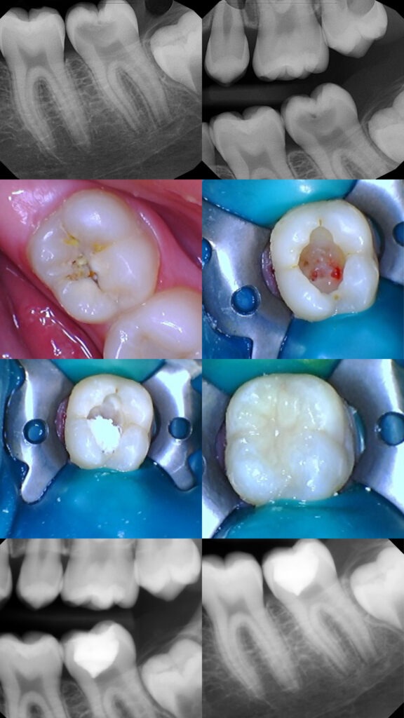
Case 1: VPT on a Carious Exposed Pulp:
A 12-year-old female presented with her mother for root canal therapy of tooth #18, with a chief complaint of mild discomfort on the lower left. Discomfort was mostly triggered by cold temperatures and sweets. The tooth responded to vitality testing, with no lingering discomfort. Percussion testing was normal. Clinically, occlusal soft gross decay was detected by tactile sensation with a restorative explorer. Radiographically, one can visualize extensive decay which appears contiguous with the pulp. The mesial and distal root apices were not fully formed. A diagnosis of reversible pulpitis with normal apical tissues was made. Due to the age of the patient, immature roots, and the pulpal/periapical diagnosis, it was decided that we would attempt to avoid full endodontic treatment at this time and would pursue caries excavation with likely pulp exposure and vital pulp therapy as the treatment of choice. If this were an older patient with mature apices, root canal therapy would have likely been performed. The patient was anesthetized, and rubber dam isolation was achieved. A high-speed diamond bur with copious water was used for initial caries penetration. A Cariosectomy Munce Bur #6 (CJM Engineering) was used to remove the bulk of the decay, and a spoon excavator was used in the deepest portion of the cavity. Once all decay was removed, a carious pulp exposure of the mesial pulp horn was encountered, with hyperemia noted. To better visualize tissue health and achieve hemostasis, a sterile #2 round carbide bur was used to remove the coronal 1-2 mm of the pulp (shallow partial pulpotomy). A lidocaine/epinephrine-soaked pellet was placed onto the pulpal wound, applying pressure for 3-5 minutes. Once hemostasis was achieved, BC RRM (Brasseler USA) was placed directly on the pulpal wound. The Munce bur, in a careful brushing motion, was used to remove excess putty from the cavity floor. The enamel and dentin were then etched with 35% phosphoric acid (Ultra-Etch, Ultradent), adhesive (Peak Universal Bond, Ultradent) was placed with a micro-brush, scrubbed onto the enamel and dentin, carefully air dried, and light cured. The tooth was then incrementally restored with shade A2 composite (Venus Diamond, Kulzer). Occlusion was checked. The patient presented for a 6-month recall and reported the tooth being asymptomatic since the procedure. Vitality testing was within normal limits. The patient was advised that close follow up would be necessary to continue monitoring the pulpal and periapical status of the tooth.
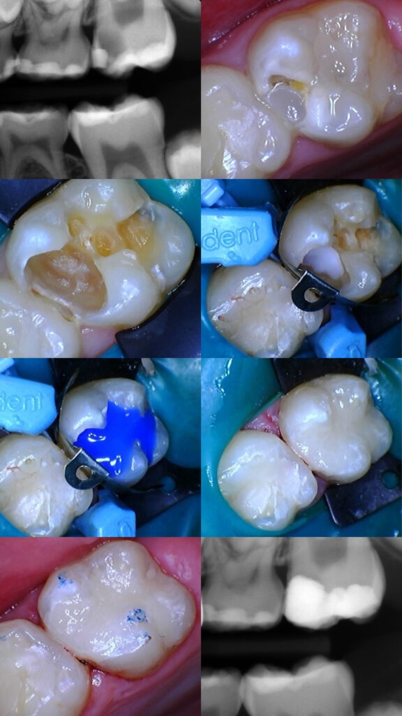
Case 2: SCR/IPC on a Deep Carious Lesion:
A 9-year male presented with his mother for root canal therapy of tooth #14. The patient reported being completely asymptomatic. Vitality testing was within normal limits. Clinically, one can visualize a broken composite on the mesial with significant recurrent decay. Radiographically, deep decay was noted extending 3/4 of the way towards the pulp. Due to the patient’s age and lack of symptoms, it was decided to do SCR/IPC, rather than full pulpectomy. If the roots were fully developed, the likely treatment would have been root canal therapy. The patient was anesthetized, and rubber dam isolation was achieved. A high-speed diamond bur with copious water was used for initial caries penetration. A Cariosectomy Munce Bur #6 (CJM Engineering) was used to remove the bulk of the decay, and a spoon excavator was used in the deepest portion of the cavity. To avoid pulp exposure, leathery/’affected’ dentin was left in closest proximity to the pulp. Care was taken to make sure the DEJ was completed cleaned to allow for sufficient bonding of the final restoration. A Lime-Lite liner (Pulpdent) was placed directly on top of the ‘affected’ dentin and light cured for 20 seconds. The enamel and dentin were then etched with 35% phosphoric acid (Ultra-Etch, Ultradent), adhesive (Peak Universal Bond, Ultradent) was placed with a micro-brush, scrubbed onto the enamel and dentin, carefully air dried, and light cured. A Palodent sectional matrix (Dentsply) was used to build up the mesial marginal ridge. The tooth was then incrementally restored with shade A2 composite (Venus Diamond, Kulzer). Occlusion was checked. The patient presented for a 6-month recall and reported the tooth being asymptomatic since the procedure. Vitality testing was within normal limits. The patient was advised that close follow up would be necessary to continue monitoring the pulpal and periapical status of the tooth.
Conclusion
The question remains regarding SCR, as to whether the pulp will succumb to the persisting dentinal infection by undergoing infectious necrosis, and how long the process might take. The fate of the dental pulp is dependent upon the number of remnant bacteria, their virulence, as well as the host resistance. If the coronal restoration remains intact, remnant bacteria may not have sufficient space to propagate to the numbers that are required to trigger infection. Conversely, pulpal necrosis may occur if the host’s resistance diminishes or if the bacteria-tight seal is compromised (15). Regarding SCR, the patient usually remains asymptomatic, and the pulp continues to test vital, even though we may see histological signs of inflammation in the pulp, clearly indicating that some remaining bacteria will not necessarily irreversibly damage the pulp tissue, at least not initially. Whether the sub-clinical inflammation in the pulp will eventually lead to irreversible infection, has not been substantiated yet. The long- term effects on the pulp, of leaving ‘affected’ dentin behind, remains unknown.
Reference:
- Aguilar P, Linsuwanont P. Vital pulp therapy in vital permanent teeth with cariously exposed pulp: a systematic review. J Endod. 2011 May;37(5):581-7. doi: 10.1016/j.joen.2010.12.004. Epub 2011 Mar 5. PMID: 21496652.
- AAE Position Statement on Vital Pulp Therapy. J Endod. 2021 Sep;47(9):1340-1344. doi: 10.1016/j.joen.2021.07.015. Epub 2021 Aug 3. PMID: 34352305
- Schwendicke F, Frencken JE, Bjørndal L, Maltz M, Manton DJ, Ricketts D, Van Landuyt K, Banerjee A, Campus G, Doméjean S, Fontana M, Leal S, Lo E, Machiulskiene V, Schulte A, Splieth C, Zandona AF, Innes NP. Managing Carious Lesions: Consensus Recommendations on Carious Tissue Removal. Adv Dent Res. 2016 May;28(2):58-67. doi: 10.1177/0022034516639271. PMID: 27099358.
- European Society of Endodontology (ESE) developed by: Duncan HF, Galler KM, Tomson PL, Simon S, El-Karim I, Kundzina R, Krastl G, Dammaschke T, Fransson H, Markvart M, Zehnder M, Bjørndal L. European Society of Endodontology position statement: Management of deep caries and the exposed pulp. Int Endod J. 2019 Jul;52(7):923-934. doi: 10.1111/iej.13080. PMID: 30664240.
- Banerjee A, Frencken JE, Schwendicke F, Innes NPT. Contemporary operative caries management: consensus recommendations on minimally invasive caries removal. Br Dent J. 2017 Aug 11;223(3):215-222. doi: 10.1038/sj.bdj.2017.672. PMID: 28798430.
- Maltz M, de Oliveira EF, Fontanella V, Bianchi R: A clinical, microbiologic, and radiographic study of deep caries lesions after incomplete caries removal. Quintessence Int 2002;33:151– 159.
- Mertz-Fairhurst EJ, Curtis JW, Ergle JW, Ruegge- berg FA, Adair SM: Ultraconservative and cariostatic sealed restorations. J Am Dent Assoc 1998;129:55–66
- BjørndalL, LarsenT: Changes in the cultivable flora in deep carious lesions following a stepwise excavation procedure. Caries Res 2000;34: 502–508.
- Bjørndal L, Larsen T, Thylstrup A: A clinical and microbiological study of deep carious lesions during stepwise excavation using long treatment intervals. Caries Res 1997;31:411–417.
- Bjørndal L, Mjör IA: Pulp-dentin biology in restorative dentistry. 4. Dental caries – Characteristics of lesions and pulpal reactions. Quintessence Int 2001;32:717–736.
- Bjørndal L, Thylstrup A: A practice-based study of stepwise excavation of deep carious lesions in permanent teeth: A 1-year follow-up study. Community Dent Oral Epidemiol 1998;26: 122–128
- Bjørndal L, Fransson H, Bruun G et al. (2017) Randomized clinical trials on deep carious lesions: 5-year follow-up. Journal of Dental Research 96, 747–53.
- Baraka M, Tekeya M, Bakry NS, Fontana M. Twelve-month randomized controlled trial of 38% silver diamine fluoride with or without potassium iodide in indirect pulp capping of young permanent molars. J Am Dent Assoc. 2022 Dec;153(12):1121-1133.e1. doi: 10.1016/j.adaj.2022.08.008. Epub 2022 Oct 15. PMID: 36253166.
- Maury Massler, Jack Pawlak, The affected and infected pulp, Oral Surgery, Oral Medicine, Oral Pathology, Volume 43, Issue 6, 1977
- Ricucci D, Siqueira JF Jr, Ro^ças IN, et al. Pulp and dentine responses to selective caries excavation: a histological and histobacteriological human study. J Dent 2020;100:103430.
- Ricucci D, Siqueira JF Jr. Bacteriologic status of non-cavitated proximal enamel caries lesions. a histologic and histobacteriologic study. J Dent 2020;100:103422
- Langeland K. Management of the inflamed pulp associated with deep carious lesion. J Endod. 1981 Apr;7(4):169-81. doi: 10.1016/S0099-2399(81)80231-2. PMID: 6939782.
- Bjørndal, S. Simon, P.L. Tomson, H.F. Duncan, Management of deep caries and the exposed pulp, Int. Endod. J. 52 (2019) 949–973, https://doi.org/10.1111/iej. 13128.
- Thompson, R.G. Craig, F.A. Curro, W.S. Green, J.A. Ship, Treatment of deep carious lesions by complete excavation or partial removal: a critical review, J. Am. Dent. Assoc. 139 (2008) 705–712, https://doi.org/10.14219/jada.archive.2008. 0252.
- A. Kidd, How “clean” must a cavity be before restoration? Caries Res. 38 (2004) 305–313, https://doi.org/10.1159/000077770.)
- Ricketts D, Lamont T, Innes NP, Kidd E, Clarkson JE. Operative caries management in adults and children. Cochrane Database Syst Rev. 2013 Mar 28;(3):CD003808. doi: 10.1002/14651858.CD003808.pub3. Update in: Cochrane Database Syst Rev. 2019 Jul 24;7:CD003808. PMID: 23543523.
- Maltz M, Garcia R, Jardim JJ, de Paula LM, Yamaguti PM, Moura MS, Garcia F, Nascimento C, Oliveira A, Mestrinho HD. Randomized trial of partial vs. stepwise caries removal: 3-year follow-up. J Dent Res. 2012 Nov;91(11):1026-31. doi: 10.1177/0022034512460403. Epub 2012 Sep 14. PMID: 22983407.
- Bjørndal L. Reentry may not be needed after partial caries removal in mainly young permanent molars with caries involving half or more of the dentin thickness. J Evid Based Dent Pract. 2013 Jun;13(2):62-3. doi: 10.1016/j.jebdp.2013.04.008. PMID: 23773470.
- Maltz M, Jardim JJ, Mestrinho HD, Yamaguti PM, Podestá K, Moura MS, de Paula LM. Partial removal of carious dentine: a multicenter randomized controlled trial and 18-month follow-up results. Caries Res. 2013;47(2):103-9. doi: 10.1159/000344013. Epub 2012 Nov 28. PMID: 23207420.
- Maltz M, Koppe B, Jardim JJ, Alves LS, de Paula LM, Yamaguti PM, Almeida JCF, Moura MS, Mestrinho HD. Partial caries removal in deep caries lesions: a 5-year multicenter randomized controlled trial. Clin Oral Investig. 2018 Apr;22(3):1337-1343. doi: 10.1007/s00784-017-2221-0. Epub 2017 Oct 8. PMID: 28988345.
- Schwendicke F, Meyer-Lueckel H, Dörfer C, Paris S. Failure of incompletely excavated teeth–a systematic review. J Dent. 2013 Jul;41(7):569-80. doi: 10.1016/j.jdent.2013.05.004. Epub 2013 May 15. PMID: 23685036.
- Bjørndal L, Larsen T, Thylstrup A. A clinical and microbiological study of deep carious lesions during stepwise excavation using long treatment intervals. Caries Res. 1997;31(6):411-7. doi: 10.1159/000262431. PMID: 9353579.
- Bjørndal L, Larsen T. Changes in the cultivable flora in deep carious lesions following a stepwise excavation procedure. Caries Res. 2000 Nov-Dec;34(6):502-8. doi: 10.1159/000016631. PMID: 11093026
- Yoshiyama M, Tay FR, Torii Y, et al. Resin adhesion to carious dentin. Am J Dent 2003;16:47–52).
- Nakajima M, Ogata M, Okuda M, Tagami J, Sano H, Pashley DH. Bonding to caries-affected dentin using self-etching primers. Am J Dent. 1999 Dec;12(6):309-14. Erratum in: Am J Dent 2000 Apr;13(2):72. PMID: 10850253. ) .
- Wei S, Sadr A, Shimada Y, Tagami J. Effect of caries-affected dentin hardness on the shear bond strength of current adhesives. J Adhes Dent. 2008;10(6):431-440.)
- Masatoshi N, Sitthikorn K, Taweesak P, Junji T. Bonding to caries-affected dentin. Jap Dent Sci Review. 2011;47(2):102-114.
- Pereira MA, Santos-Júnior RBD, Tavares JA, Oliveira AH, Leal PC, Takeshita WM, Barbosa-Júnior AM, Bertassoni LEB, Faria-E-Silva AL. No additional benefit of using a calcium hydroxide liner during stepwise caries removal: A randomized clinical trial. J Am Dent Assoc. 2017 Jun;148(6):369-376. doi: 10.1016/j.adaj.2017.02.019. Epub 2017 Mar 23. PMID: 28343596.
- Baraka M, Tekeya M, Bakry NS, Fontana M. Twelve-month randomized controlled trial of 38% silver diamine fluoride with or without potassium iodide in indirect pulp capping of young permanent molars. J Am Dent Assoc. 2022 Dec;153(12):1121-1133.e1. doi: 10.1016/j.adaj.2022.08.008. Epub 2022 Oct 15. PMID: 36253166.
- Kotsanos N, Arizos S. Evaluation of a resin modified glass ionomer serving both as indirect pulp therapy and as restorative material for primary molars. Eur Arch Paediatr Dent. 2011 Jun;12(3):170-5. doi: 10.1007/BF03262801. PMID: 21640064.
- Costa CA, Giro EM, Nascimento AB, et al. Short-term evaluation of the pulpo-dentin complex response to a resin-modified glass-ionomer cement and a bonding agent applied in deep cavities. Dent Mater. 2003;19(8):739-746.
- de Souza Costa CA, Teixeira HM, Lopes do Nascimento AB, Hebling J. Biocompatibility of resin-based dental materials applied as liners in deep cavities prepared in human teeth. J Biomed Mater Res B Appl Biomater. 2007;81(1):175-184.
- Alex G. Adhesive considerations in the placement of direct composite restorations. Functional Esthetics and Restorative Dentistry. 2007;1(1):20-25.
- Sidhu SK, Schmalz G. The biocompatibility of glass- ionomer cement materials. A status report for the American Journal of Dentistry. Am J Dent 2001: 14: 387–396.
- Tarim B, Hafez AA, Cox CF. Pulpal response to a resin- modified glass-ionomer material on non-exposed and exposed monkey pulps. Quintessence Int 1998: 29: 535–542
- Murray P, Hafez A, Windsor L, Smith A, Cox C. Comparison of pulp responses following restoration of exposed and non-exposed cavities. Journal of Dentistry 2002;30:213–222. [PubMed: 12450712]
- Heys R, Fitzgerald M. Microleakage of three cement bases. Journal of Dental Research 1991;70(1):55–58. [PubMed: 1991861]
- Murray P, Hafez A, Smith A, Cox C. Bacterial microleakage and pulp inflammation associated with various restorative materials. Dental Materials 2002;18:470–478. [PubMed: 12098576]
- de Souza Costa C, Giro E, Lopes do Nascimento A, Teixeira H, Hebling J. Short-term evaluation of the pulpodentin complex response to a resin-modified glass-ionomer cement and a bonding agent applied in deep cavities. Dental Materials 2003;19:739–746. [PubMed: 14511732]
- Barros MMAF, De Queiroz Rodrigues MI, Muniz FWMG, Rodrigues LKA. Selective, stepwise, or nonselective removal of carious tissue: which technique offers lower risk for the treatment of dental caries in permanent teeth? A systematic review and meta-analysis. Clin Oral Investig. 2020 Feb;24(2):521-532. doi: 10.1007/s00784-019-03114-5. Epub 2019 Nov 26. PMID: 31773371.
- Jardim JJ, Mestrinho HD, Koppe B, de Paula LM, Alves LS, Yamaguti PM, Almeida JCF, Maltz M. Restorations after selective caries removal: 5-Year randomized trial. J Dent. 2020 Aug;99:103416. doi: 10.1016/j.jdent.2020.103416. Epub 2020 Jun 22. PMID: 32585263.
- Li T, Zhai X, Song F, Zhu H. Selective versus non-selective removal for dental caries: a systematic review and meta-analysis. Acta Odontol Scand. 2018 Mar;76(2):135-140. doi: 10.1080/00016357.2017.1392602. Epub 2017 Oct 26. PMID: 29073814.)
- Ricketts D. Deep or partial caries removal: which is best? Evid Based Dent. 2008;9(3):71-2. doi: 10.1038/sj.ebd.6400592. PMID: 18927562.
- Uribe S. Partial caries removal in symptomless teeth reduces the risk of pulp exposure. Evid Based Dent. 2006;7(4):94. doi: 10.1038/sj.ebd.6400444. PMID: 17187037.
- Ricketts DN, Kidd EA, Innes N, Clarkson J. Complete or ultraconservative removal of decayed tissue in unfilled teeth. Cochrane Database Syst Rev. 2006 Jul 19;(3):CD003808. doi: 10.1002/14651858.CD003808.pub2. Update in: Cochrane Database Syst Rev. 2013;3:CD003808. PMID: 16856019.
- Thompson V, Craig RG, Curro FA, Green WS, Ship JA. Treatment of deep carious lesions by complete excavation or partial removal: a critical review. J Am Dent Assoc. 2008 Jun;139(6):705-12. doi: 10.14219/jada.archive.2008.0252. PMID: 18519994; PMCID: PMC2692285
- Matsuo T, Nakanishi T, Shimizu H, et al. A clinical study of direct pulp capping applied to carious-exposed pulps. J Endod 1996;22:551.